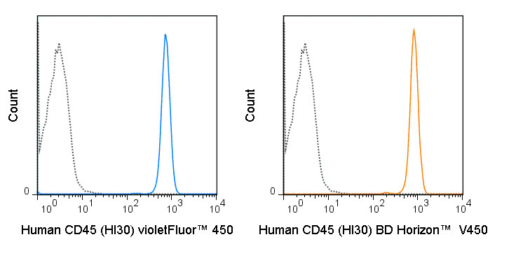
violetFluor™ 450 Anti-Human CD45 (HI30) Antibody
- SPECIFICATION
- CITATIONS
- PROTOCOLS
- BACKGROUND

Application
| FC |
|---|---|
| Isotype | Mouse IgG1, kappa |
| Concentration | 5 uL (1.0 ug)/test |
| Reactivity | Human |
| Formulation | 10 mM NaH2PO4, 150 mM NaCl, 0.09% NaN3, 0.1% gelatin, pH7.2 |
| Gene ID | 5788 |
|---|---|
| Gene Name | PTPRC |
| Alternative Name(s) | Leukocyte Common Antigen, LCA, Ly-5 |
| Format | violetFluor™ 450 |
| Preparation | This monoclonal antibody was purified from tissue culture supernatant via affinity chromatography. The purified antibody was conjugated under optimal conditions, with unreacted dye removed from the preparation. It is recommended to store the product undiluted at 4°C, and protected from prolonged exposure to light. Do not freeze. |
| Application Notes | This antibody preparation has been pre-titrated and quality-tested for flow cytometry using an appropriate cell type. The antibody has been diluted for use at 5 uL per test, defined as the amount of antibody that will stain a cell sample in a final volume of approximately 100 uL. The number of cells within a sample should be determined empirically, but typically ranges between 1x10e5 to 1x10e8 cells. violetFluor™ 450 dye is excited by the violet (405 nm) laser and has a peak emission of 450 nm. The most common band pass filters for this dye are 440/40 or 450/50. violetFluor™ 450 can be used as an alternative for Pacific Blue®, BD Horizon™ V450 or eFluor® 450. |
| Storage Conditions | 2-8°C protected from light |
Strowig T, Rongvaux A, Rathinam C, Takizawa H, Borsotti C, Philbrick W, Eynon EE, Manz MG, and Flavell RA. 2011. Proc. Natl. Acad. Sci. 108: 13218-13223. (Flow Cytometry)
Kim M-H, Suh H-S, Si Q, Terman BE, and Lee SC. 2006. J. Virol. 80: 62-72. (in vitro blocking, Western Blot)
Zhang M and Varki A. 2004. Glycobiology. 14: 939-949. (Immunoprecipitation)
Gelbmann CM, Leeb SN, Vogl D, Maendel M, Herfarth H, Scholmerich J, Falk W, and Rogler G. 2003. Gut. 52:1448-1456. (Immunocytochemistry)
Yamada T, Zhu D, Saxon A, and Zhang K. 2002. J. Biol. Chem. 277(32): 28830-28835. (in vitro blocking)
Esser MT, Graham DR, Coren LV, Trubey CM, Bess JW, Arthur LO, Ott DE, and Lifson JD. 2001. J. Virol. 75(13)6173-6182. (Western Blot)
Goto E, Kohrogi H, Hirata N, Tsumori K, Hirosako S, Hamamoto J, Fujii K, Kawano O, and Ando M. 2000. Am. J. Respir. Cell Mol. Biol. 22: 405. (Immunohistochemistry – frozen tissue)
Esser MT, Graham DR, Coren LV, Trubey CM, Bess JW, Arthur LO, Ott DE, and Lifson JD. 2001. J. Virol. 75(13)6173-6182. (Western Blot)
Provided below are standard protocols that you may find useful for product applications.
Background
The HI30 antibody reacts with human CD45, one of the most abundant hematopoietic markers and one that is expressed on all leukocytes (the Leukocyte Common Antigen, LCA). CD45 is a protein tyrosine phosphatase existing in several isoforms, each being generated and expressed in cell-specific patterns. With its broad cell distribution, CD45 is critical for many leukocyte functions, regulating signal transduction and cell activation associated with the T cell receptor, B cell receptor, and IL-2 receptor. Other forms of CD45, with restricted cellular expression, include CD45R (B220), CD45RA, CD45RB, CD45RO and others.
The HI30 antibody is widely used as a marker for human CD45 expression on T cells, B cells, monocytes, macrophages, and NK cells.
If you have used an Abcepta product and would like to share how it has performed, please click on the "Submit Review" button and provide the requested information. Our staff will examine and post your review and contact you if needed.
If you have any additional inquiries please email technical services at tech@abcepta.com.


 Foundational characteristics of cancer include proliferation, angiogenesis, migration, evasion of apoptosis, and cellular immortality. Find key markers for these cellular processes and antibodies to detect them.
 The SUMOplot™ Analysis Program predicts and scores sumoylation sites in your protein. SUMOylation is a post-translational modification involved in various cellular processes, such as nuclear-cytosolic transport, transcriptional regulation, apoptosis, protein stability, response to stress, and progression through the cell cycle.
 The Autophagy Receptor Motif Plotter predicts and scores autophagy receptor binding sites in your protein. Identifying proteins connected to this pathway is critical to understanding the role of autophagy in physiological as well as pathological processes such as development, differentiation, neurodegenerative diseases, stress, infection, and cancer.


