


 Foundational characteristics of cancer include proliferation, angiogenesis, migration, evasion of apoptosis, and cellular immortality. Find key markers for these cellular processes and antibodies to detect them.
 The SUMOplot™ Analysis Program predicts and scores sumoylation sites in your protein. SUMOylation is a post-translational modification involved in various cellular processes, such as nuclear-cytosolic transport, transcriptional regulation, apoptosis, protein stability, response to stress, and progression through the cell cycle.
 The Autophagy Receptor Motif Plotter predicts and scores autophagy receptor binding sites in your protein. Identifying proteins connected to this pathway is critical to understanding the role of autophagy in physiological as well as pathological processes such as development, differentiation, neurodegenerative diseases, stress, infection, and cancer.
Anti-PIAS1 (RABBIT) Antibody
PIAS1 Antibody
- SPECIFICATION
- CITATIONS
- PROTOCOLS
- BACKGROUND
| Host | Rabbit |
|---|---|
| Conjugate | Unconjugated |
| Target Species | Human |
| Reactivity | Human |
| Clonality | Polyclonal |
Application
| WB, E, I, LCI |
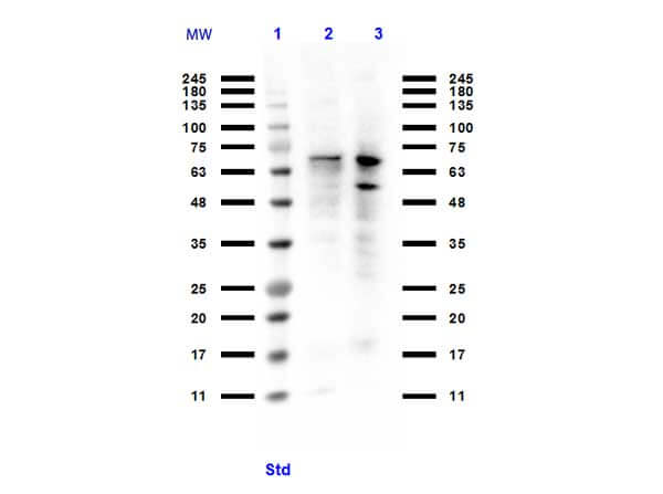
| Application Note | Anti-PIAS1 Antibody has been tested for use in ELISA, Western Blotting, and Dot Blotting. HEK293 WCL p/n (W09-000-365) and K-562 WCL p/n (W09-001-GJ7) were used as positive control lysates in western blot. Expect a band at approximately 72 kDa in Western Blots of specific cell lysates and tissues. Specific conditions for reactivity should be optimized by the end user. While not tested, it is likely Anti-PIAS1 antibody will work for immunofluorescence, IHC, and CHIP assays. |
| Physical State | Liquid (sterile filtered) |
| Buffer | 0.01 M Sodium Phosphate, 0.25 M Sodium Chloride, pH 7.2 |
| Immunogen | Anti-PIAS1 antibody was prepared from whole rabbit serum produced by repeated immunizations with a synthetic peptide from the internal region of human PIAS1. |
| Preservative | 0.01% (w/v) Sodium Azide |
| Gene ID | 8554 |
|---|---|
| Other Names | 8554 |
| Purity | Anti-PIAS1 Antibody was affinity purified from monospecific antiserum by immunoaffinity chromatography. A BLAST analysis was used to suggest reactivity to mouse 92.9% homology for the immunogen sequence. Cross-reactivity from PIAS1 family and other sources has not been determined. |
| Storage Condition | Store vial at -20° C prior to opening. Aliquot contents and freeze at -20° C or below for extended storage. Avoid cycles of freezing and thawing. Centrifuge product if not completely clear after standing at room temperature. This product is stable for several weeks at 4° C as an undiluted liquid. Dilute only prior to immediate use. |
| Precautions Note | This product is for research use only and is not intended for therapeutic or diagnostic applications. |
| Name | PIAS1 |
|---|---|
| Synonyms | DDXBP1 |
| Function | Functions as an E3-type small ubiquitin-like modifier (SUMO) ligase, stabilizing the interaction between UBE2I and the substrate, and as a SUMO-tethering factor (PubMed:11583632, PubMed:11867732, PubMed:14500712, PubMed:21965678, PubMed:36050397). Catalyzes sumoylation of various proteins, such as CEBPB, MRE11, MTA1, PTK2 and PML (PubMed:11583632, PubMed:11867732, PubMed:14500712, PubMed:21965678, PubMed:36050397). Plays a crucial role as a transcriptional coregulation in various cellular pathways, including the STAT pathway, the p53 pathway and the steroid hormone signaling pathway (PubMed:11583632, PubMed:11867732). In vitro, binds A/T-rich DNA (PubMed:15133049). The effects of this transcriptional coregulation, transactivation or silencing, may vary depending upon the biological context (PubMed:11583632, PubMed:11867732, PubMed:14500712, PubMed:21965678, PubMed:36050397). Mediates sumoylation of MRE11, stabilizing MRE11 on chromatin during end resection (PubMed:36050397). Sumoylates PML (at 'Lys-65' and 'Lys-160') and PML-RAR and promotes their ubiquitin-mediated degradation (By similarity). PIAS1-mediated sumoylation of PML promotes its interaction with CSNK2A1/CK2 which in turn promotes PML phosphorylation and degradation (By similarity). Enhances the sumoylation of MTA1 and may participate in its paralog- selective sumoylation (PubMed:21965678). Plays a dynamic role in adipogenesis by promoting the SUMOylation and degradation of CEBPB (By similarity). Mediates the nuclear mobility and localization of MSX1 to the nuclear periphery, whereby MSX1 is brought into the proximity of target myoblast differentiation factor genes (By similarity). Also required for the binding of MSX1 to the core enhancer region in target gene promoter regions, independent of its sumoylation activity (By similarity). Capable of binding to the core enhancer region TAAT box in the MYOD1 gene promoter (By similarity). |
| Cellular Location | Nucleus {ECO:0000250|UniProtKB:O88907}. Nucleus speckle Nucleus, PML body {ECO:0000250|UniProtKB:O88907}. Cytoplasm, cytoskeleton. Note=Interaction with CSRP2 may induce a partial redistribution along the cytoskeleton (PubMed:11672422). Interaction with MSX1 is required for localization to the nuclear periphery (By similarity) {ECO:0000250|UniProtKB:O88907, ECO:0000269|PubMed:11672422} |
| Tissue Location | Expressed in numerous tissues with highest level in testis. |

Thousands of laboratories across the world have published research that depended on the performance of antibodies from Abcepta to advance their research. Check out links to articles that cite our products in major peer-reviewed journals, organized by research category.
info@abcepta.com, and receive a free "I Love Antibodies" mug.

Provided below are standard protocols that you may find useful for product applications.
Background
The PIAS proteins (protein inhibitor of activated STAT) play a crucial role as transcriptional coregulators in various cellular pathways, including the STAT, p53 and the steroid hormone signaling pathway. The PIAS protein family includes at least five evolutionarily conserved genes, including PIAS1. The major function of the PIAS proteins is the control of gene transcription and can also act as small ubiquitin-like-modifier (SUMO) E3 ligases. PIAS1 binds specifically to STAT1, inhibiting STAT1-mediated gene activation and also binds to the Gu/RNA helicase II enzyme, leading to the proteolytic cleavage of Gu/RH-II. PIAS1 is a potent co-activator for CP2c-mediated alpha-globin expression in erythroid cells. Anti-PIAS1 Antibody is useful for researchers interested in epigenetics and cancer research.
If you have used an Abcepta product and would like to share how it has performed, please click on the "Submit Review" button and provide the requested information. Our staff will examine and post your review and contact you if needed.
If you have any additional inquiries please email technical services at tech@abcepta.com.
Ordering Information
Other Products
Shipping Information
