


 Foundational characteristics of cancer include proliferation, angiogenesis, migration, evasion of apoptosis, and cellular immortality. Find key markers for these cellular processes and antibodies to detect them.
 The SUMOplot™ Analysis Program predicts and scores sumoylation sites in your protein. SUMOylation is a post-translational modification involved in various cellular processes, such as nuclear-cytosolic transport, transcriptional regulation, apoptosis, protein stability, response to stress, and progression through the cell cycle.
 The Autophagy Receptor Motif Plotter predicts and scores autophagy receptor binding sites in your protein. Identifying proteins connected to this pathway is critical to understanding the role of autophagy in physiological as well as pathological processes such as development, differentiation, neurodegenerative diseases, stress, infection, and cancer.
Anti-Fibroblast Activation Protein (FAP) (RABBIT) Antibody
Fibroblast Activation Protein Antibody
- SPECIFICATION
- CITATIONS
- PROTOCOLS
- BACKGROUND

| Host | Rabbit |
|---|---|
| Conjugate | Unconjugated |
| Target Species | Mouse |
| Reactivity | Mouse |
| Clonality | Polyclonal |
Application
| WB, E, I, LCI |
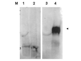
| Application Note | This affinity purified antibody has been tested for use in ELISA and western blotting. Specific conditions for reactivity should be optimized by the end user. Expect a band approximately 88 kDa in size corresponding to FAP by western blotting in the appropriate cell lysate or extract. |
| Physical State | Liquid (sterile filtered) |
| Buffer | 0.02 M Potassium Phosphate, 0.15 M Sodium Chloride, pH 7.2 |
| Immunogen | This affinity purified antibody was prepared from whole rabbit serum produced by repeated immunizations with a synthetic peptide corresponding to an internal region of mouse FAP protein. |
| Preservative | 0.01% (w/v) Sodium Azide |
| Gene ID | 14089 |
|---|---|
| Other Names | 14089 |
| Purity | This product was affinity purified from monospecific antiserum by immunoaffinity chromatography. This antibody is known to react with mouse FAP protein. A BLAST analysis was used to suggest partial cross-reactivity with FAP from human and rat sources based on ~88% homology with the immunizing sequence. Reactivity with FAP from other sources has not been determined. |
| Storage Condition | Store vial at -20° C prior to opening. Aliquot contents and freeze at -20° C or below for extended storage. Avoid cycles of freezing and thawing. Centrifuge product if not completely clear after standing at room temperature. This product is stable for several weeks at 4° C as an undiluted liquid. Dilute only prior to immediate use. |
| Precautions Note | This product is for research use only and is not intended for therapeutic or diagnostic applications. |
| Name | Fap {ECO:0000312|MGI:MGI:109608} |
|---|---|
| Function | Cell surface glycoprotein serine protease that participates in extracellular matrix degradation and involved in many cellular processes including tissue remodeling, fibrosis, wound healing, inflammation and tumor growth. Both plasma membrane and soluble forms exhibit post-proline cleaving endopeptidase activity, with a marked preference for Ala/Ser-Gly-Pro-Ser/Asn/Ala consensus sequences, on substrate such as alpha-2-antiplasmin SERPINF2 and SPRY2. Degrade also gelatin, heat-denatured type I collagen, but not native collagen type I and IV, vibronectin, tenascin, laminin, fibronectin, fibrin or casein. Also has dipeptidyl peptidase activity, exhibiting the ability to hydrolyze the prolyl bond two residues from the N-terminus of synthetic dipeptide substrates provided that the penultimate residue is proline, with a preference for Ala-Pro, Ile-Pro, Gly-Pro, Arg-Pro and Pro-Pro. Natural neuropeptide hormones for dipeptidyl peptidase are the neuropeptide Y (NPY), peptide YY (PYY), substance P (TAC1) and brain natriuretic peptide 32 (NPPB). The plasma membrane form, in association with either DPP4, PLAUR or integrins, is involved in the pericellular proteolysis of the extracellular matrix (ECM), and hence promotes cell adhesion, migration and invasion through the ECM. Plays a role in tissue remodeling during development and wound healing. Participates in the cell invasiveness towards the ECM in malignant melanoma cancers. Enhances tumor growth progression by increasing angiogenesis, collagen fiber degradation and apoptosis and by reducing antitumor response of the immune system. Promotes glioma cell invasion through the brain parenchyma by degrading the proteoglycan brevican. Acts as a tumor suppressor in melanocytic cells through regulation of cell proliferation and survival in a serine protease activity-independent manner. |
| Cellular Location | [Prolyl endopeptidase FAP]: Cell surface. Cell membrane {ECO:0000250|UniProtKB:Q12884}; Single-pass type II membrane protein. Cell projection, lamellipodium membrane {ECO:0000250|UniProtKB:Q12884}; Single-pass type II membrane protein. Cell projection, invadopodium membrane {ECO:0000250|UniProtKB:Q12884}; Single-pass type II membrane protein. Cell projection, ruffle membrane {ECO:0000250|UniProtKB:Q12884}; Single-pass type II membrane protein. Membrane {ECO:0000250|UniProtKB:Q12884}; Single-pass type II membrane protein. Note=Localized on cell surface with lamellipodia and invadopodia membranes and on shed vesicles Colocalized with DPP4 at invadopodia and lamellipodia membranes of migratory activated endothelial cells in collagenous matrix Colocalized with DPP4 on endothelial cells of capillary-like microvessels but not large vessels within invasive breast ductal carcinoma. Anchored and enriched preferentially by integrin alpha- 3/beta-1 at invadopodia, plasma membrane protrusions that correspond to sites of cell invasion, in a collagen-dependent manner. Localized at plasma and ruffle membranes in a collagen-independent manner Colocalized with PLAUR preferentially at the cell surface of invadopodia membranes in a cytoskeleton-, integrin- and vitronectin- dependent manner. Concentrated at invadopodia membranes, specialized protrusions of the ventral plasma membrane in a fibrobectin-dependent manner. Colocalizes with extracellular components (ECM), such as collagen fibers and fibronectin. {ECO:0000250|UniProtKB:Q12884} |
| Tissue Location | Expressed strongly in uterus, pancreas, submaxillary gland and skin, less in lymph node, ovary, skeletal muscle, adrenal and bone marrow. Expressed in reactive stromal fibroblast in epithelial cancers. Expressed in melanocytes but not melanomas (at protein level). Detected in fibroblasts, in placenta, uterus, embryos from day 7-19 and in newborn mice (P1) |

Thousands of laboratories across the world have published research that depended on the performance of antibodies from Abcepta to advance their research. Check out links to articles that cite our products in major peer-reviewed journals, organized by research category.
info@abcepta.com, and receive a free "I Love Antibodies" mug.

Provided below are standard protocols that you may find useful for product applications.
Background
This antibody is designed, produced, and validated as part of a collaboration between Rockland and the National Cancer Institute (NCI) and is suitable for Cancer, Immunology and Nuclear Signaling research. Fibroblast Activation Protein (FAP) is expressed in the stroma of sites that are undergoing wound healing. In addition, it has recently been reported that FAP is expressed in the stroma of sites of metastatic disease. Inhibition of FAP may lead to a dramatic decrease in the number of metastatic osteosarcoma lung nodules. FAP exists as an inactive monomer and when activated forms homodimers or heterodimers with DPP4. Multiple isoforms of FAP are reported as alternative splicing products from a common gene.
If you have used an Abcepta product and would like to share how it has performed, please click on the "Submit Review" button and provide the requested information. Our staff will examine and post your review and contact you if needed.
If you have any additional inquiries please email technical services at tech@abcepta.com.
Ordering Information
Other Products
Shipping Information
