Anti-c-MET pY1349 pY1356 (RABBIT) Antibody
c-Met phospho Y1349 / pY1356 Antibody
- SPECIFICATION
- CITATIONS
- PROTOCOLS
- BACKGROUND

| Host | Rabbit |
|---|---|
| Conjugate | Unconjugated |
| Target Species | Human |
| Reactivity | Human |
| Clonality | Polyclonal |
Application
| WB, E, I, LCI |
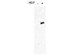
| Application Note | This affinity purified antibody has been tested for use in ELISA and western blot. Specific conditions for reactivity should be optimized by the end user. Expect a band approximately 150 kDa in size corresponding to phosphorylated c-Met protein by western blotting in the appropriate cell lysate or extract. This phospho-specific polyclonal antibody reacts with human c-Met pY1349pY1356 and shows minimal reactivity by ELISA against the non-phosphorylated form of the immunizing peptide. |
| Physical State | Liquid (sterile filtered) |
| Buffer | 0.02 M Potassium Phosphate, 0.15 M Sodium Chloride, pH 7.2 |
| Immunogen | This affinity purified antibody was prepared from whole rabbit serum produced by repeated immunizations with a synthetic peptide corresponding to residues surrounding Y1349 and Y1356 of human c-Met protein. |
| Preservative | 0.01% (w/v) Sodium Azide |
| Gene ID | 4233 |
|---|---|
| Other Names | 4233 |
| Purity | Phospho c-MET Antibody was produced from monospecific antiserum by immunoaffinity chromatography using dual-phospho-peptide coupled to agarose beads followed by cross-adsorption against nonphospho-peptide. Antibody is specific for human c-Met protein phosphorylated at Y1349 and Y1356. A BLAST analysis was used to suggest cross-reactivity with c-Met from human, mouse and rat based on 100% homology with the immunizing sequence. Cross-reactivity with c-Met from other sources has not been determined. |
| Storage Condition | Store Phospho antibody at -20° C prior to opening. Aliquot contents and freeze at -20° C or below for extended storage. Avoid cycles of freezing and thawing. Centrifuge product if not completely clear after standing at room temperature. This product is stable for several weeks at 4° C as an undiluted liquid. Dilute only prior to immediate use. |
| Precautions Note | This product is for research use only and is not intended for therapeutic or diagnostic applications. |
| Name | MET |
|---|---|
| Function | Receptor tyrosine kinase that transduces signals from the extracellular matrix into the cytoplasm by binding to hepatocyte growth factor/HGF ligand. Regulates many physiological processes including proliferation, scattering, morphogenesis and survival. Ligand binding at the cell surface induces autophosphorylation of MET on its intracellular domain that provides docking sites for downstream signaling molecules. Following activation by ligand, interacts with the PI3-kinase subunit PIK3R1, PLCG1, SRC, GRB2, STAT3 or the adapter GAB1. Recruitment of these downstream effectors by MET leads to the activation of several signaling cascades including the RAS-ERK, PI3 kinase-AKT, or PLCgamma-PKC. The RAS-ERK activation is associated with the morphogenetic effects while PI3K/AKT coordinates prosurvival effects. During embryonic development, MET signaling plays a role in gastrulation, development and migration of neuronal precursors, angiogenesis and kidney formation. During skeletal muscle development, it is crucial for the migration of muscle progenitor cells and for the proliferation of secondary myoblasts (By similarity). In adults, participates in wound healing as well as organ regeneration and tissue remodeling. Promotes also differentiation and proliferation of hematopoietic cells. May regulate cortical bone osteogenesis (By similarity). |
| Cellular Location | Membrane; Single-pass type I membrane protein. |
| Tissue Location | Expressed in normal hepatocytes as well as in epithelial cells lining the stomach, the small and the large intestine Found also in basal keratinocytes of esophagus and skin. High levels are found in liver, gastrointestinal tract, thyroid and kidney. Also present in the brain. Expressed in metaphyseal bone (at protein level) (PubMed:26637977). |

Thousands of laboratories across the world have published research that depended on the performance of antibodies from Abcepta to advance their research. Check out links to articles that cite our products in major peer-reviewed journals, organized by research category.
info@abcepta.com, and receive a free "I Love Antibodies" mug.
Provided below are standard protocols that you may find useful for product applications.
Background
This antibody is designed, produced, and validated as part of a collaboration between Rockland and the National Cancer Institute (NCI) and is suitable for Cancer, Immunology and Nuclear Signaling research. Anti-c-MET is the receptor for hepatocyte growth factor (also known as scatter factor, HGF/SF), and belongs to the tyrosine kinase superfamily. Interaction of c-Met with HGF results in autophosphorylation of c-Met at multiple tyrosines. Phosphorylation of Y1234/1235 in the c-Met kinase domain is critical to kinase activation. When phosphorylated, Y1349 and Y1356, along with surrounding amino acids, form a unique bidentate docking site for substrates such as Gab1, Grb2, phosphatidylinositol 3-kinase (PI3K) and others. C-Met mainly uses the Gab1 scaffolding adaptor in its initial step of signal transmission. Well-characterized downstream signaling pathways that are activated by c-Met include the ERK/MAPK, PI3K–Akt/PKB, Crk–Rap and Rac–Pak pathways, resulting in proliferation and increased cell survival. Anti-Phospho cMET was developed with NCI and is ideal for Cancer Research.
If you have used an Abcepta product and would like to share how it has performed, please click on the "Submit Review" button and provide the requested information. Our staff will examine and post your review and contact you if needed.
If you have any additional inquiries please email technical services at tech@abcepta.com.


 Foundational characteristics of cancer include proliferation, angiogenesis, migration, evasion of apoptosis, and cellular immortality. Find key markers for these cellular processes and antibodies to detect them.
 The SUMOplot™ Analysis Program predicts and scores sumoylation sites in your protein. SUMOylation is a post-translational modification involved in various cellular processes, such as nuclear-cytosolic transport, transcriptional regulation, apoptosis, protein stability, response to stress, and progression through the cell cycle.
 The Autophagy Receptor Motif Plotter predicts and scores autophagy receptor binding sites in your protein. Identifying proteins connected to this pathway is critical to understanding the role of autophagy in physiological as well as pathological processes such as development, differentiation, neurodegenerative diseases, stress, infection, and cancer.


