Anti-HER3 (RABBIT) Antibody
HER3 Antibody
- SPECIFICATION
- CITATIONS
- PROTOCOLS
- BACKGROUND

| Host | Rabbit |
|---|---|
| Conjugate | Unconjugated |
| Target Species | Human |
| Reactivity | Human |
| Clonality | Polyclonal |
Application
| WB, E, IP, I, LCI |
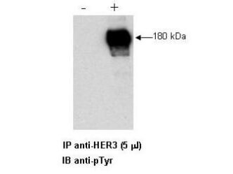
| Application Note | Anti-HER3 antibody has been tested by ELISA, immunoprecipitation, and western blot and is specifically designed for immunoprecipitation and can be used for immunoblotting in conjunction with a detection antibody like anti-phosphotyrosine. Reactivity in other assays is likely, but has not been determined. Recognition of HER3 is independent of the phosphorylation status of the molecule. Human prostate cell lines (LNCap) or human breast cell line (MDA-453) are typically used as positive control sources. For immunoprecipitation use approximately 5 µL of the antibody. The immunoprecipitation mix contained the antibody, 25 µL of Protein A-agarose beads and 1.0 ml of lysate (lysate contains approximately 1.0 mg of total protein). This mixture is rotated overnight at 4°C and then washed 3 times with lysis buffer (used to prepare the lysate). The resulting bead complex is dissolved in 20-30 µL of 3X SDS-PAGE sample buffer and approximately 15 µL is loaded per lane on an 8% polyacrylamide gel. The combination of immunoprecipitation and immunoblotting was performing using the anti-HER3 antibody for immunoprecipitation followed by immunoblot detection using an anti-phosphotyrosine antibody. The complex is then reacted with HRP Goat-anti-Rabbit IgG and ECL for detection and shows a single HER3 band at 180kDa. Researchers should determine optimal titers for other applications. |
| Physical State | Liquid (sterile filtered) |
| Buffer | 0.02 M Potassium Phosphate, 0.15 M Sodium Chloride, pH 7.2 |
| Immunogen | Anti-HER3 whole rabbit serum was prepared by repeated immunizations with a HER3 fusion protein corresponding to amino acids 1283 to 1323 (40 amino acids) at the carboxy-terminus of human HER3. |
| Preservative | 0.01% (w/v) Sodium Azide |
| Gene ID | 2065 |
|---|---|
| Other Names | 2065 |
| Purity | Rabbit Anti-HER3 is an IgG fraction antibody purified from monospecific antiserum by Protein A chromatography followed by cross adsorption against GST and extensive dialysis against the buffer stated above. Assay by immunoelectrophoresis resulted in a single precipitin arc against anti-Rabbit Serum. The antibody is directed against human HER3 and is useful in determining its presence in immunoprecipitation experiments. This antibody can detect HER3 from human, mouse and rat sources. Reactivity of this antibody with HER3 from other species is unknown. |
| Storage Condition | Store Anti-HER3 at -20° C prior to opening. Aliquot contents and freeze at -20° C or below for extended storage. Avoid cycles of freezing and thawing. Centrifuge product if not completely clear after standing at room temperature. This product is stable for several weeks at 4° C as an undiluted liquid. Dilute only prior to immediate use. |
| Precautions Note | This product is for research use only and is not intended for therapeutic or diagnostic applications. |
| Name | ERBB3 |
|---|---|
| Synonyms | HER3 |
| Function | Tyrosine-protein kinase that plays an essential role as cell surface receptor for neuregulins. Binds to neuregulin-1 (NRG1) and is activated by it; ligand-binding increases phosphorylation on tyrosine residues and promotes its association with the p85 subunit of phosphatidylinositol 3-kinase (PubMed:20682778). May also be activated by CSPG5 (PubMed:15358134). Involved in the regulation of myeloid cell differentiation (PubMed:27416908). |
| Cellular Location | [Isoform 1]: Cell membrane; Single-pass type I membrane protein |
| Tissue Location | Epithelial tissues and brain. |

Thousands of laboratories across the world have published research that depended on the performance of antibodies from Abcepta to advance their research. Check out links to articles that cite our products in major peer-reviewed journals, organized by research category.
info@abcepta.com, and receive a free "I Love Antibodies" mug.
Provided below are standard protocols that you may find useful for product applications.
Background
HER3 antibody is ideal for western blotting, ELISA and IP. The human Epidermal Growth Factor Receptor (EGFR) family is a family of receptor tyrosine kinases consisting of 4 members: EGFR, HER2, HER3 and HER4. These receptors are expressed on a wide variety of tissue types and have been implicated in many human cancers, including breast, gastric, colon and prostate. Strong expression of both HER3 and HER2 (also known as p185neu) by tumor cells can be associated with poor outcome for patients. The ligand for HER3 and HER4 is called Neu Differentiation Factor (NDF) or Heregulin. In some instances, antibodies against these receptors prove effective in combination therapy regimens for treatment of the disease. Signaling through the EGFR family involves the combinatorial dimerization of the four known family members and is mediated by at least 8 different hormones, including EGF and the heregulins. Heregulin binding and tyrosine phosphorylation by HER3 or HER4 is enhanced in the presence of HER2. Structurally the members of the EGFR family are 1210 to 1342 amino acids in length with approximately 650 amino acids of the extracellular domain being heavily glycosylated. The extracellular domains show up to 70% sequence homology.
If you have used an Abcepta product and would like to share how it has performed, please click on the "Submit Review" button and provide the requested information. Our staff will examine and post your review and contact you if needed.
If you have any additional inquiries please email technical services at tech@abcepta.com.


 Foundational characteristics of cancer include proliferation, angiogenesis, migration, evasion of apoptosis, and cellular immortality. Find key markers for these cellular processes and antibodies to detect them.
 The SUMOplot™ Analysis Program predicts and scores sumoylation sites in your protein. SUMOylation is a post-translational modification involved in various cellular processes, such as nuclear-cytosolic transport, transcriptional regulation, apoptosis, protein stability, response to stress, and progression through the cell cycle.
 The Autophagy Receptor Motif Plotter predicts and scores autophagy receptor binding sites in your protein. Identifying proteins connected to this pathway is critical to understanding the role of autophagy in physiological as well as pathological processes such as development, differentiation, neurodegenerative diseases, stress, infection, and cancer.


