


 Foundational characteristics of cancer include proliferation, angiogenesis, migration, evasion of apoptosis, and cellular immortality. Find key markers for these cellular processes and antibodies to detect them.
 The SUMOplot™ Analysis Program predicts and scores sumoylation sites in your protein. SUMOylation is a post-translational modification involved in various cellular processes, such as nuclear-cytosolic transport, transcriptional regulation, apoptosis, protein stability, response to stress, and progression through the cell cycle.
 The Autophagy Receptor Motif Plotter predicts and scores autophagy receptor binding sites in your protein. Identifying proteins connected to this pathway is critical to understanding the role of autophagy in physiological as well as pathological processes such as development, differentiation, neurodegenerative diseases, stress, infection, and cancer.
Anti-HSP27 (MOUSE) Monoclonal Antibody
HSP27 Antibody
- SPECIFICATION
- CITATIONS
- PROTOCOLS
- BACKGROUND

| Host | Mouse |
|---|---|
| Conjugate | Unconjugated |
| Target Species | Human |
| Reactivity | Human |
| Clonality | Monoclonal |
Application
| WB, E, I, LCI |
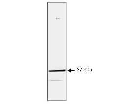
| Application Note | This protein A purified monoclonal antibody against human Hsp27 has been tested for use in immunoblotting and ELISA and is suitable in immunoprecipitation, immunohistochemistry, and immunocytochemistry. The antibody recognizes a 27 kDa band corresponding to hsp27 in cell lysates from breast carcinoma. Both frozen sections and paraffin embedded material can be used for immunocytochemistry and immunohistochemistry. |
| Physical State | Liquid (sterile filtered) |
| Buffer | 0.02 M Potassium Phosphate, 0.5 M Sodium Chloride, pH 7.2 |
| Immunogen | This HSP27 monoclonal antibody was produced by repeated immunizations with a prokaryotic recombinant protein corresponding to the full length human hsp27 protein. |
| Preservative | 0.01% (w/v) Sodium Azide |
| Gene ID | 3315 |
|---|---|
| Other Names | 3315 |
| Purity | This protein A purified mouse monoclonal antibody reacts specifically with HSP27 in human tissues and cell lines. MCF-7 cells are recommended as a positive control. Cross reactivity with hsp27 from other mammalian sources is likely. No cross reactivity occurs with HSP70, HSP90 or HSP104. |
| Storage Condition | Store vial at -20° C prior to opening. Aliquot contents and freeze at -20° C or below for extended storage. Avoid cycles of freezing and thawing. Centrifuge product if not completely clear after standing at room temperature. This product is stable for several weeks at 4° C as an undiluted liquid. Dilute only prior to immediate use. |
| Precautions Note | This product is for research use only and is not intended for therapeutic or diagnostic applications. |
| Name | HSPB1 |
|---|---|
| Synonyms | HSP27, HSP28 |
| Function | Small heat shock protein which functions as a molecular chaperone probably maintaining denatured proteins in a folding- competent state (PubMed:10383393, PubMed:20178975). Plays a role in stress resistance and actin organization (PubMed:19166925). Through its molecular chaperone activity may regulate numerous biological processes including the phosphorylation and the axonal transport of neurofilament proteins (PubMed:23728742). |
| Cellular Location | Cytoplasm. Nucleus Cytoplasm, cytoskeleton, spindle Note=Cytoplasmic in interphase cells. Colocalizes with mitotic spindles in mitotic cells. Translocates to the nucleus during heat shock and resides in sub-nuclear structures known as SC35 speckles or nuclear splicing speckles. |
| Tissue Location | Detected in all tissues tested: skeletal muscle, heart, aorta, large intestine, small intestine, stomach, esophagus, bladder, adrenal gland, thyroid, pancreas, testis, adipose tissue, kidney, liver, spleen, cerebral cortex, blood serum and cerebrospinal fluid. Highest levels are found in the heart and in tissues composed of striated and smooth muscle. |

Thousands of laboratories across the world have published research that depended on the performance of antibodies from Abcepta to advance their research. Check out links to articles that cite our products in major peer-reviewed journals, organized by research category.
info@abcepta.com, and receive a free "I Love Antibodies" mug.

Provided below are standard protocols that you may find useful for product applications.
Background
Heat shock protein (HSP) 27 is one of the small HSPs that are constitutively expressed at different levels in different cell types and tissues (this protein has also been referred to as the Estrogen-Regulated 24 kDa protein, hsp25 and hsp28). Like other small heat shock proteins, HSP27 is regulated at both the transcriptional and post-translational level. In response to stress, the expression level of HSP27 increases several-fold to confer cellular resistance to the adverse environmental change. The common functions of sHsps are chaperone activity, thermotolerance, inhibition of apoptosis, regulation of cell development, and cell differentiation. They also take part in signal transduction. The HSP27 gene has 3 exons. The mouse Hsp25 gene was mapped to chromosome 5 in a region homologous to 7q in the human. They also mapped the mouse Hsp105 gene to chromosome 5 but suggested that the human homolog is probably on 13q, not chromosome 7. HSP27 plays a major role in the increased thermal resistance acquired by cells after exposure to HSP inducers. The level of HSP27 phosphorylation is significantly elevated after exposure of cells to heat shock, sodium arsenite, IL-1 and TNF-a. MAPKAPK2 and MAPKAPK3 are both activated by these conditions and can phosphorylate HSP27 on serine residues Anti-HSP27 Antibody is ideal for investigators involved in Signaling Proteins, Cell Stress & Chaperone Proteins, Cancer, Cellular Stress, and p38 Pathway research.
If you have used an Abcepta product and would like to share how it has performed, please click on the "Submit Review" button and provide the requested information. Our staff will examine and post your review and contact you if needed.
If you have any additional inquiries please email technical services at tech@abcepta.com.
Ordering Information
Other Products
Shipping Information
