Anti-Hif-1α hydroxy P564 (RABBIT) Antibody
HIF-1-alpha hydroxy P564 Antibody
- SPECIFICATION
- CITATIONS
- PROTOCOLS
- BACKGROUND

| Host | Rabbit |
|---|---|
| Conjugate | Unconjugated |
| Target Species | Human |
| Reactivity | Human |
| Clonality | Polyclonal |
Application
| WB, IHC, E, I, LCI |
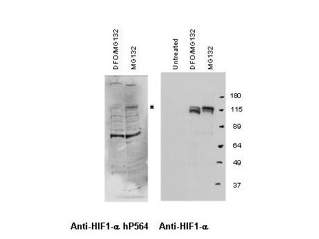
| Application Note | This antibody has been tested for use in ELISA and western blotting. Specific conditions for reactivity should be optimized by the end user. Expect a band approximately 110 kDa in size corresponding to HIF-1a hydroxyl P564 by western blotting in the appropriate cell lysate or extract. |
| Physical State | Liquid (sterile filtered) |
| Buffer | 0.02 M Potassium Phosphate, 0.15 M Sodium Chloride, pH 7.2 |
| Immunogen | This antibody was prepared from whole rabbit serum produced by repeated immunizations with a synthetic peptide corresponding to a region surrounding the P564 of human HIF-1a. |
| Preservative | 0.01% (w/v) Sodium Azide |
| Gene ID | 3091 |
|---|---|
| Other Names | 3091 |
| Purity | This antibody is directed against human HIF-1a hydroxyP564 and is specific for the hydroxylated form of the protein. Minimal reactivity occurs with the non-hydroxylated form of the protein. This antibody is specific for HIF-1a hydroxylated at P564. Minimal cross-reactivity occurs with non-hydroxylated HIF-1a. A BLAST analysis was used to suggest cross-reactivity with HIF-1a from human, monkey, mouse, rat, dog, bovine and Xenopus sources based on a 100% homology with the immunizing sequence. Reactivity against homologues from other sources is not known. |
| Storage Condition | Store vial at -20° C prior to opening. Aliquot contents and freeze at -20° C or below for extended storage. Avoid cycles of freezing and thawing. Centrifuge product if not completely clear after standing at room temperature. This product is stable for several weeks at 4° C as an undiluted liquid. Dilute only prior to immediate use. |
| Precautions Note | This product is for research use only and is not intended for therapeutic or diagnostic applications. |
| Name | HIF1A {ECO:0000303|PubMed:7539918, ECO:0000312|HGNC:HGNC:4910} |
|---|---|
| Function | Functions as a master transcriptional regulator of the adaptive response to hypoxia (PubMed:11292861, PubMed:11566883, PubMed:15465032, PubMed:16973622, PubMed:17610843, PubMed:18658046, PubMed:20624928, PubMed:22009797, PubMed:30125331, PubMed:9887100). Under hypoxic conditions, activates the transcription of over 40 genes, including erythropoietin, glucose transporters, glycolytic enzymes, vascular endothelial growth factor, HILPDA, and other genes whose protein products increase oxygen delivery or facilitate metabolic adaptation to hypoxia (PubMed:11292861, PubMed:11566883, PubMed:15465032, PubMed:16973622, PubMed:17610843, PubMed:20624928, PubMed:22009797, PubMed:30125331, PubMed:9887100). Plays an essential role in embryonic vascularization, tumor angiogenesis and pathophysiology of ischemic disease (PubMed:22009797). Heterodimerizes with ARNT; heterodimer binds to core DNA sequence 5'-TACGTG-3' within the hypoxia response element (HRE) of target gene promoters (By similarity). Activation requires recruitment of transcriptional coactivators such as CREBBP and EP300 (PubMed:16543236, PubMed:9887100). Activity is enhanced by interaction with NCOA1 and/or NCOA2 (PubMed:10594042). Interaction with redox regulatory protein APEX1 seems to activate CTAD and potentiates activation by NCOA1 and CREBBP (PubMed:10202154, PubMed:10594042). Involved in the axonal distribution and transport of mitochondria in neurons during hypoxia (PubMed:19528298). |
| Cellular Location | Cytoplasm. Nucleus. Nucleus speckle {ECO:0000250|UniProtKB:Q61221}. Note=Colocalizes with HIF3A in the nucleus and speckles (By similarity). Cytoplasmic in normoxia, nuclear translocation in response to hypoxia (PubMed:9822602) {ECO:0000250|UniProtKB:Q61221, ECO:0000269|PubMed:9822602} |
| Tissue Location | Expressed in most tissues with highest levels in kidney and heart. Overexpressed in the majority of common human cancers and their metastases, due to the presence of intratumoral hypoxia and as a result of mutations in genes encoding oncoproteins and tumor suppressors. A higher level expression seen in pituitary tumors as compared to the pituitary gland. |

Thousands of laboratories across the world have published research that depended on the performance of antibodies from Abcepta to advance their research. Check out links to articles that cite our products in major peer-reviewed journals, organized by research category.
info@abcepta.com, and receive a free "I Love Antibodies" mug.
Provided below are standard protocols that you may find useful for product applications.
Background
This antibody is designed, produced, and validated as part of a collaboration between Rockland and the National Cancer Institute (NCI) and is suitable for Cancer, Immunology and Nuclear Signaling research. Tumor hypoxia often directly correlates with aggressive phenotype, metastasis progression and resistance to chemotherapy. HIF-1 transcription factors are dramatically induced in hypoxic areas and regulate the expression of genes necessary for tumor adaptation to conditions of low oxygen. The stabilization of HIF-1a by hypoxia is critically dependent upon the hydroxylation of certain Proline residues that exist in the oxygen-dependent degradation domain of HIF-1a. HIF factors are now considered an important therapeutic target for cancer intervention. HIF-1a is useful to researchers interested in cell metabolism, cell survival, and angiogenesis.
If you have used an Abcepta product and would like to share how it has performed, please click on the "Submit Review" button and provide the requested information. Our staff will examine and post your review and contact you if needed.
If you have any additional inquiries please email technical services at tech@abcepta.com.


 Foundational characteristics of cancer include proliferation, angiogenesis, migration, evasion of apoptosis, and cellular immortality. Find key markers for these cellular processes and antibodies to detect them.
 The SUMOplot™ Analysis Program predicts and scores sumoylation sites in your protein. SUMOylation is a post-translational modification involved in various cellular processes, such as nuclear-cytosolic transport, transcriptional regulation, apoptosis, protein stability, response to stress, and progression through the cell cycle.
 The Autophagy Receptor Motif Plotter predicts and scores autophagy receptor binding sites in your protein. Identifying proteins connected to this pathway is critical to understanding the role of autophagy in physiological as well as pathological processes such as development, differentiation, neurodegenerative diseases, stress, infection, and cancer.


