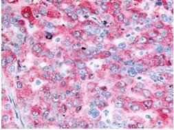
Anti-EGFR (RABBIT) Antibody
EGFR Antibody
- SPECIFICATION
- CITATIONS
- PROTOCOLS
- BACKGROUND

| Host | Rabbit |
|---|---|
| Conjugate | Unconjugated |
| Target Species | Human |
| Reactivity | Rat, Human |
| Clonality | Polyclonal |
Application
| WB, IHC, E, IP, I, LCI |
| Application Note | Anti-EGFR antibody has been tested by and is specifically designed for ELISA, immunoblotting, immunoprecipitation, and immunohistochemistry. Reactivity in other assays is likely, but has not been determined. Recognition of EGFR is independent of the phosphorylation status at tyrosine 1173. A431 cells, keratinocytes in normal epidermis, or placenta are typically used as positive control sources. The antigen is typically localized in the cell membrane. For western blotting, good results are also achieved on PVDF membranes blocked with 5% lowfat milk diluted in TTBS for 1 hour at room temperature. Also, dilute the primary antibody and secondary in 5% lowfat milk in TTBS. Anti-EGFR can be diluted up to 1:10,000 for immunoblot depending on the cell line and the amount of EGFR in a particular lysate. For immunoprecipitation, use approximately 10 µl of the antibody. The immunoprecipitation mix should contain the antibody, 25 µl of Protein A-agarose beads and 1.0 ml of lysate (lysate contains approximately 1.0 mg of total protein). This mixture should be rotated overnight at 4°C and then washed 3 times with lysis buffer (used to prepare the lysate). The resulting bead complex is dissolved in 20-30 µl of 3X SDS-PAGE sample buffer and approximately 15 µl is loaded per lane on an 8% polyacrylamide gel. |
| Physical State | Liquid (sterile filtered) |
| Immunogen | This whole rabbit serum was prepared by repeated immunizations with a peptide synthesized using conventional technology. The sequence of the epitope maps to a region near the carboxy terminus which is identical in human, mouse and rat EGFR. |
| Preservative | 0.01% (w/v) Sodium Azide |
| Gene ID | 1956 |
|---|---|
| Other Names | 1956 |
| Purity | This antiserum is directed against human epidermal growth factor receptor (EGFR) and is useful in determining its presence in western blotting and immunoprecipitation experiments. This antibody can detect EGFR from human, mouse and rat sources. Reactivity of this antibody with EGFR from other species is unknown. No reaction is observed against ErbB-2, ErbB-3 or ErbB-4. |
| Storage Condition | Store vial at -20° C prior to opening. Aliquot contents and freeze at -20° C or below for extended storage. Avoid cycles of freezing and thawing. Centrifuge product if not completely clear after standing at room temperature. This product is stable for several weeks at 4° C as an undiluted liquid. Dilute only prior to immediate use. |
| Precautions Note | This product is for research use only and is not intended for therapeutic or diagnostic applications. |
| Name | EGFR (HGNC:3236) |
|---|---|
| Synonyms | ERBB, ERBB1, HER1 |
| Function | Receptor tyrosine kinase binding ligands of the EGF family and activating several signaling cascades to convert extracellular cues into appropriate cellular responses (PubMed:10805725, PubMed:27153536, PubMed:2790960, PubMed:35538033). Known ligands include EGF, TGFA/TGF- alpha, AREG, epigen/EPGN, BTC/betacellulin, epiregulin/EREG and HBEGF/heparin-binding EGF (PubMed:12297049, PubMed:15611079, PubMed:17909029, PubMed:20837704, PubMed:27153536, PubMed:2790960, PubMed:7679104, PubMed:8144591, PubMed:9419975). Ligand binding triggers receptor homo- and/or heterodimerization and autophosphorylation on key cytoplasmic residues. The phosphorylated receptor recruits adapter proteins like GRB2 which in turn activates complex downstream signaling cascades. Activates at least 4 major downstream signaling cascades including the RAS-RAF-MEK-ERK, PI3 kinase-AKT, PLCgamma-PKC and STATs modules (PubMed:27153536). May also activate the NF-kappa-B signaling cascade (PubMed:11116146). Also directly phosphorylates other proteins like RGS16, activating its GTPase activity and probably coupling the EGF receptor signaling to the G protein-coupled receptor signaling (PubMed:11602604). Also phosphorylates MUC1 and increases its interaction with SRC and CTNNB1/beta-catenin (PubMed:11483589). Positively regulates cell migration via interaction with CCDC88A/GIV which retains EGFR at the cell membrane following ligand stimulation, promoting EGFR signaling which triggers cell migration (PubMed:20462955). Plays a role in enhancing learning and memory performance (By similarity). Plays a role in mammalian pain signaling (long-lasting hypersensitivity) (By similarity). |
| Cellular Location | Cell membrane; Single-pass type I membrane protein. Endoplasmic reticulum membrane; Single-pass type I membrane protein Golgi apparatus membrane; Single-pass type I membrane protein. Nucleus membrane; Single-pass type I membrane protein. Endosome. Endosome membrane. Nucleus. Note=In response to EGF, translocated from the cell membrane to the nucleus via Golgi and ER (PubMed:17909029, PubMed:20674546). Endocytosed upon activation by ligand (PubMed:17182860, PubMed:17909029, PubMed:27153536, PubMed:2790960). Colocalized with GPER1 in the nucleus of estrogen agonist-induced cancer-associated fibroblasts (CAF) (PubMed:20551055) |
| Tissue Location | Ubiquitously expressed. Isoform 2 is also expressed in ovarian cancers. |

Thousands of laboratories across the world have published research that depended on the performance of antibodies from Abcepta to advance their research. Check out links to articles that cite our products in major peer-reviewed journals, organized by research category.
info@abcepta.com, and receive a free "I Love Antibodies" mug.
Provided below are standard protocols that you may find useful for product applications.
Background
EGFR is a transmembrane glycoprotein that is a member of a family of protein tyrosine kinases crucial to maintaining a normal balance in cell growth and development. Growth factor receptors are involved not only in promoting the proliferation of normal cells but also in the aberrant growth of many types of human tumors. For example, the epidermal growth factor receptor (EGFR) is mutated and/or over-expressed in many common solid human squamous cell carcinomas including breast, brain, bladder, lung, gastric, head & neck, esophagus, cervix, vulva, ovary, and endometrium. Over-expression of the EGFR gene occurs in carcinomas with and without gene amplification. EGFR and ErbB-2 are particularly important in breast cancer because increased production or activation has been associated with poor prognosis. EGFR belongs to a family of growth factor receptors, which also includes ErbB-2/HER-2/neu, ErbB-3/HER-3/neu and ErbB-4/HER-4/neu. EGFR can heterodimerize with each of the members of this family.
If you have used an Abcepta product and would like to share how it has performed, please click on the "Submit Review" button and provide the requested information. Our staff will examine and post your review and contact you if needed.
If you have any additional inquiries please email technical services at tech@abcepta.com.


 Foundational characteristics of cancer include proliferation, angiogenesis, migration, evasion of apoptosis, and cellular immortality. Find key markers for these cellular processes and antibodies to detect them.
 The SUMOplot™ Analysis Program predicts and scores sumoylation sites in your protein. SUMOylation is a post-translational modification involved in various cellular processes, such as nuclear-cytosolic transport, transcriptional regulation, apoptosis, protein stability, response to stress, and progression through the cell cycle.
 The Autophagy Receptor Motif Plotter predicts and scores autophagy receptor binding sites in your protein. Identifying proteins connected to this pathway is critical to understanding the role of autophagy in physiological as well as pathological processes such as development, differentiation, neurodegenerative diseases, stress, infection, and cancer.


