


 Foundational characteristics of cancer include proliferation, angiogenesis, migration, evasion of apoptosis, and cellular immortality. Find key markers for these cellular processes and antibodies to detect them.
 The SUMOplot™ Analysis Program predicts and scores sumoylation sites in your protein. SUMOylation is a post-translational modification involved in various cellular processes, such as nuclear-cytosolic transport, transcriptional regulation, apoptosis, protein stability, response to stress, and progression through the cell cycle.
 The Autophagy Receptor Motif Plotter predicts and scores autophagy receptor binding sites in your protein. Identifying proteins connected to this pathway is critical to understanding the role of autophagy in physiological as well as pathological processes such as development, differentiation, neurodegenerative diseases, stress, infection, and cancer.
TAK1 Antibody
Rabbit Anti-Human TAK1 Polyclonal
- SPECIFICATION
- CITATIONS
- PROTOCOLS
- BACKGROUND

Application
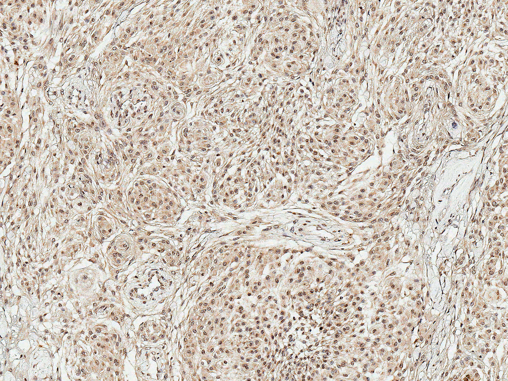
| IHC |
|---|---|
| Primary Accession | O43318 |
| Other Accession | NP_003179.1 |
| Host | Rabbit |
| Reactivity | Rat |
| Clonality | Polyclonal |
| Format | TAK1 |
| Target/Specificity | TAK1 |
| Other Names | MAP3K7 Antibody, TAK1 Antibody, Mitogen-activated protein kinase kinase kinase 7 Antibody, TGF-beta-activated kinase 1 Antibody, Transforming growth factor-beta-activated kinase 1 Antibody, MEKK7 Antibody |
| Clone Names | TAK1 |
| Immunogen | Synthetic peptide of Human TAK1 (400-500 aa), conjugated to Keyhole Limpet Haemocyanin (KLH). |
| Purification | Peptide Affinity Purified |
| Storage | -20ºC |
| Storage Buffer | PBS pH 7.4, 50% glycerol, 0.09% sodium azide *Storage buffer may change when conjugated |
| Shipping Temperature | Blue Ice or 4ºC |
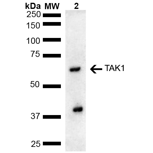
| Certificate of Analysis | A 1:1000 dilution of SPC-736 was sufficient for detection of TAK1 in 15 µg of rat liver cell lysates by ECL immunoblot analysis using goat anti-rabbit IgG:HRP as the secondary antibody. |
| Cellular Localization | Cytoplasm | Cell Membrane | Peripheral Membrane Protein | Cytoplasmic Side |

Thousands of laboratories across the world have published research that depended on the performance of antibodies from Abcepta to advance their research. Check out links to articles that cite our products in major peer-reviewed journals, organized by research category.
info@abcepta.com, and receive a free "I Love Antibodies" mug.

Provided below are standard protocols that you may find useful for product applications.
Background
TAK1 encoded by the gene MAP3K7, is a protein-serine/threonine kinase that is activated by proinflammatory cytokines and in response to physical/chemical stress, including UVR, osmotic- and oxidative stress. It is a mediator of TRAF6 and TGF-β signal transduction, and activates IKBKB and MAPK8 in response to TRAF6 signalling. It also stimulates NFκB activation and activation of the p38 MAPK pathway. It is responsible for controlling a variety of cell functions such as transcription and apoptosis. TAK1 is important for TGF-β1 regulation of MMP9 and the metastatic potential of certain breast cancer cell lines.
If you have used an Abcepta product and would like to share how it has performed, please click on the "Submit Review" button and provide the requested information. Our staff will examine and post your review and contact you if needed.
If you have any additional inquiries please email technical services at tech@abcepta.com.
Ordering Information
Other Products
Shipping Information
