


 Foundational characteristics of cancer include proliferation, angiogenesis, migration, evasion of apoptosis, and cellular immortality. Find key markers for these cellular processes and antibodies to detect them.
 The SUMOplot™ Analysis Program predicts and scores sumoylation sites in your protein. SUMOylation is a post-translational modification involved in various cellular processes, such as nuclear-cytosolic transport, transcriptional regulation, apoptosis, protein stability, response to stress, and progression through the cell cycle.
 The Autophagy Receptor Motif Plotter predicts and scores autophagy receptor binding sites in your protein. Identifying proteins connected to this pathway is critical to understanding the role of autophagy in physiological as well as pathological processes such as development, differentiation, neurodegenerative diseases, stress, infection, and cancer.
CD98 Antibody
Rabbit mAb
- SPECIFICATION
- CITATIONS
- PROTOCOLS
- BACKGROUND

Application
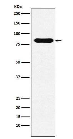
| WB, IHC, IP |
|---|---|
| Primary Accession | P08195 |
| Clonality | Monoclonal |
| Other Names | 4T2HC; CD98; CD98HC; MDU1; NACAE; Slc3a2; |
| Isotype | Rabbit IgG |
| Host | Rabbit |
| Calculated MW | 67994 Da |
| Dilution | WB 1:500~1:2000 IHC 1:50~1:200 IP 1:50 |
|---|---|
| Purification | Affinity-chromatography |
| Immunogen | A synthesized peptide derived from human CD98 |
| Description | Required for the function of light chain amino-acid transporters. Involved in sodium-independent, high-affinity transport of large neutral amino acids such as phenylalanine, tyrosine, leucine, arginine and tryptophan. Involved in guiding and targeting of LAT1 and LAT2 to the plasma membrane. |
| Storage Condition and Buffer | Rabbit IgG in phosphate buffered saline , pH 7.4, 150mM NaCl, 0.02% sodium azide and 50% glycerol. Store at +4°C short term. Store at -20°C long term. Avoid freeze / thaw cycle. |
| Name | SLC3A2 (HGNC:11026) |
|---|---|
| Synonyms | MDU1 |
| Function | Acts as a chaperone that facilitates biogenesis and trafficking of functional transporters heterodimers to the plasma membrane. Forms heterodimer with SLC7 family transporters (SLC7A5, SLC7A6, SLC7A7, SLC7A8, SLC7A10 and SLC7A11), a group of amino-acid antiporters (PubMed:10574970, PubMed:10903140, PubMed:11557028, PubMed:30867591, PubMed:33298890, PubMed:33758168, PubMed:34880232, PubMed:9751058, PubMed:9829974, PubMed:9878049). Heterodimers function as amino acids exchangers, the specificity of the substrate depending on the SLC7A subunit. Heterodimers SLC3A2/SLC7A6 or SLC3A2/SLC7A7 mediate the uptake of dibasic amino acids (PubMed:10903140, PubMed:9829974). Heterodimer SLC3A2/SLC7A11 functions as an antiporter by mediating the exchange of extracellular anionic L-cystine and intracellular L-glutamate across the cellular plasma membrane (PubMed:34880232). SLC3A2/SLC7A10 translocates small neutral L- and D- amino acids across the plasma membrane (By similarity). SLC3A2/SLC75 or SLC3A2/SLC7A8 translocates neutral amino acids with broad specificity, thyroid hormones and L-DOPA (PubMed:10574970, PubMed:11389679, PubMed:11557028, PubMed:11564694, PubMed:11742812, PubMed:12117417, PubMed:12225859, PubMed:12716892, PubMed:15980244, PubMed:30867591, PubMed:33298890, PubMed:33758168). SLC3A2 is essential for plasma membrane localization, stability, and the transport activity of SLC7A5 and SLC7A8 (PubMed:10391915, PubMed:10574970, PubMed:11311135, PubMed:15769744, PubMed:33066406). When associated with LAPTM4B, the heterodimer SLC7A5 is recruited to lysosomes to promote leucine uptake into these organelles, and thereby mediates mTORC1 activation (PubMed:25998567). Modulates integrin-related signaling and is essential for integrin-dependent cell spreading, migration and tumor progression (PubMed:11121428, PubMed:15625115). |
| Cellular Location | Apical cell membrane. Cell membrane; Single-pass type II membrane protein. Cell junction {ECO:0000250|UniProtKB:P10852}. Lysosome membrane. Melanosome. Basolateral cell membrane {ECO:0000250|UniProtKB:P10852}. Note=Localized at the plasma membrane when associated with SLC7A5/LAT1 or SLC7A8/LAT2 (PubMed:11311135, PubMed:9751058). Localized to the apical membrane of placental syncytiotrophoblastic cells (PubMed:11742812). Recruited to lysosomes by LAPTM4B (PubMed:25998567). Identified by mass spectrometry in melanosome fractions from stage I to stage IV (PubMed:17081065) Located selectively at cell-cell adhesion sites (By similarity) Colocalized with SLC7A8/LAT2 at the basolateral membrane of kidney proximal tubules and small intestine epithelia. Expressed in both luminal and abluminal membranes of brain capillary endothelial cells (By similarity). {ECO:0000250|UniProtKB:P10852, ECO:0000269|PubMed:11311135, ECO:0000269|PubMed:11742812, ECO:0000269|PubMed:17081065, ECO:0000269|PubMed:25998567, ECO:0000269|PubMed:9751058} |
| Tissue Location | Expressed ubiquitously in all tissues tested with highest levels detected in kidney, placenta and testis and weakest level in thymus. During gestation, expression in the placenta was significantly stronger at full-term than at the mid-trimester stage Expressed in HUVECS and at low levels in resting peripheral blood T- lymphocytes and quiescent fibroblasts. Also expressed in fetal liver and in the astrocytic process of primary astrocytic gliomas. Expressed in retinal endothelial cells and in the intestinal epithelial cell line C2BBe1. |
Research Areas
Citations (0)

Thousands of laboratories across the world have published research that depended on the performance of antibodies from Abcepta to advance their research. Check out links to articles that cite our products in major peer-reviewed journals, organized by research category.
Submit your citation using an Abcepta antibody to
info@abcepta.com, and receive a free "I Love Antibodies" mug.
info@abcepta.com, and receive a free "I Love Antibodies" mug.

Application Protocols
Provided below are standard protocols that you may find useful for product applications.
Abcepta welcomes feedback from its customers.
If you have used an Abcepta product and would like to share how it has performed, please click on the "Submit Review" button and provide the requested information. Our staff will examine and post your review and contact you if needed.
If you have any additional inquiries please email technical services at tech@abcepta.com.
$ 350.00
$ 175.00
Cat# AP91865
Ordering Information
United States
AlbaniaAustraliaAustriaBelgiumBosnia & HerzegovinaBrazilBulgariaCanadaCentral AmericaChinaCroatiaCyprusCzech RepublicDenmarkEstoniaFinlandFranceGermanyGreeceHong KongHungaryIcelandIndiaIndonesiaIrelandIsraelItalyJapanLatviaLithuaniaLuxembourgMacedoniaMalaysiaMaltaNetherlandsNew ZealandNorwayPakistanPolandPortugalRomaniaSerbiaSingaporeSlovakiaSloveniaSouth AfricaSouth KoreaSpainSwedenSwitzerlandTaiwanTurkeyUnited KingdomUnited StatesVietnamWorldwideOthers
USA Headquarters
(888) 735-7227 / (858) 622-0099 or (858) 875-1900
Other Products
Shipping Information
Domestic orders (in stock items)
Shipped out the same day. Orders placed after 1 PM (PST) will ship out the next business day.
International orders
Contact your local distributors
