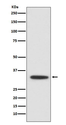
XBP1 Antibody
Rabbit mAb
- SPECIFICATION
- CITATIONS
- PROTOCOLS
- BACKGROUND

Application
| WB, IHC, FC, ICC |
|---|---|
| Primary Accession | P17861 |
| Clonality | Monoclonal |
| Other Names | TREB5; XBP1; XBP2; |
| Isotype | Rabbit IgG |
| Host | Rabbit |
| Calculated MW | 28695 Da |
| Dilution | WB 1:500~1:2000 IHC 1:100~1:500 ICC/IF 1:50~1:200 FC 1:50 |
|---|---|
| Purification | Affinity-chromatography |
| Immunogen | A synthesized peptide derived from human XBP1 |
| Description | Acts during endoplasmic reticulum stress (ER) by activating unfolded protein response (UPR) target genes via direct binding to the UPR element (UPRE). Binds DNA preferably to the CRE-like element 5'-GATGACGTG[TG]N(3)[AT]T-3', and also to some TPA response elements (TRE). Binds to the HLA DR-alpha promoter. Binds to the Tax-responsive element (TRE) of HTLV-I. |
| Storage Condition and Buffer | Rabbit IgG in phosphate buffered saline , pH 7.4, 150mM NaCl, 0.02% sodium azide and 50% glycerol. Store at +4°C short term. Store at -20°C long term. Avoid freeze / thaw cycle. |
| Name | XBP1 (HGNC:12801) |
|---|---|
| Function | Functions as a transcription factor during endoplasmic reticulum (ER) stress by regulating the unfolded protein response (UPR). Required for cardiac myogenesis and hepatogenesis during embryonic development, and the development of secretory tissues such as exocrine pancreas and salivary gland (By similarity). Involved in terminal differentiation of B lymphocytes to plasma cells and production of immunoglobulins (PubMed:11460154). Modulates the cellular response to ER stress in a PIK3R-dependent manner (PubMed:20348923). Binds to the cis-acting X box present in the promoter regions of major histocompatibility complex class II genes (PubMed:8349596). Involved in VEGF-induced endothelial cell (EC) proliferation and retinal blood vessel formation during embryonic development but also for angiogenesis in adult tissues under ischemic conditions. Functions also as a major regulator of the UPR in obesity-induced insulin resistance and type 2 diabetes for the management of obesity and diabetes prevention (By similarity). |
| Cellular Location | Endoplasmic reticulum. Note=Colocalizes with ERN1 and KDR in the endoplasmic reticulum in endothelial cells in a vascular endothelial growth factor (VEGF)-dependent manner (PubMed:23529610) [Isoform 2]: Nucleus. Cytoplasm {ECO:0000250|UniProtKB:O35426}. Note=Localizes predominantly in the nucleus. Colocalizes in the nucleus with SIRT1. Translocates into the nucleus in a PIK3R-, ER stress-induced- and/or insulin-dependent manner (By similarity). {ECO:0000250|UniProtKB:O35426} |
| Tissue Location | Expressed in plasma cells in rheumatoid synovium (PubMed:11460154). Over-expressed in primary breast cancer and metastatic breast cancer cells (PubMed:25280941). Isoform 1 and isoform 2 are expressed at higher level in proliferating as compared to confluent quiescent endothelial cells (PubMed:19416856) |

Thousands of laboratories across the world have published research that depended on the performance of antibodies from Abcepta to advance their research. Check out links to articles that cite our products in major peer-reviewed journals, organized by research category.
info@abcepta.com, and receive a free "I Love Antibodies" mug.
Provided below are standard protocols that you may find useful for product applications.
If you have used an Abcepta product and would like to share how it has performed, please click on the "Submit Review" button and provide the requested information. Our staff will examine and post your review and contact you if needed.
If you have any additional inquiries please email technical services at tech@abcepta.com.


 Foundational characteristics of cancer include proliferation, angiogenesis, migration, evasion of apoptosis, and cellular immortality. Find key markers for these cellular processes and antibodies to detect them.
 The SUMOplot™ Analysis Program predicts and scores sumoylation sites in your protein. SUMOylation is a post-translational modification involved in various cellular processes, such as nuclear-cytosolic transport, transcriptional regulation, apoptosis, protein stability, response to stress, and progression through the cell cycle.
 The Autophagy Receptor Motif Plotter predicts and scores autophagy receptor binding sites in your protein. Identifying proteins connected to this pathway is critical to understanding the role of autophagy in physiological as well as pathological processes such as development, differentiation, neurodegenerative diseases, stress, infection, and cancer.


