


 Foundational characteristics of cancer include proliferation, angiogenesis, migration, evasion of apoptosis, and cellular immortality. Find key markers for these cellular processes and antibodies to detect them.
 The SUMOplot™ Analysis Program predicts and scores sumoylation sites in your protein. SUMOylation is a post-translational modification involved in various cellular processes, such as nuclear-cytosolic transport, transcriptional regulation, apoptosis, protein stability, response to stress, and progression through the cell cycle.
 The Autophagy Receptor Motif Plotter predicts and scores autophagy receptor binding sites in your protein. Identifying proteins connected to this pathway is critical to understanding the role of autophagy in physiological as well as pathological processes such as development, differentiation, neurodegenerative diseases, stress, infection, and cancer.
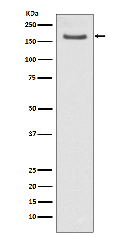
FANCD2 Antibody
Rabbit mAb
- SPECIFICATION
- CITATIONS
- PROTOCOLS
- BACKGROUND

Application
| WB, IHC, ICC, IP |
|---|---|
| Primary Accession | Q9BXW9 |
| Reactivity | Rat |
| Clonality | Monoclonal |
| Other Names | FA D2; FA4; FAC D2; FACD 2; FACD; FACD2; FAD; FAD2; FANCD 2; FANCD; FANCD2; Fanconi anemia group D2 protein; FLJ23826; Protein FACD2; Type 4 Fanconi pancytopenia; |
| Isotype | Rabbit IgG |
| Host | Rabbit |
| Calculated MW | 164128 Da |
| Dilution | WB 1:1000~1:2000 IHC 1:50~1:200 ICC/IF 1:50~1:200 IP 1:50 |
|---|---|
| Purification | Affinity-chromatography |
| Immunogen | A synthesized peptide derived from human FANCD2 |
| Description | Required for maintenance of chromosomal stability. Promotes accurate and efficient pairing of homologs during meiosis. Involved in the repair of DNA double-strand breaks, both by homologous recombination and single-strand annealing. |
| Storage Condition and Buffer | Rabbit IgG in phosphate buffered saline , pH 7.4, 150mM NaCl, 0.02% sodium azide and 50% glycerol. Store at +4°C short term. Store at -20°C long term. Avoid freeze / thaw cycle. |
| Name | FANCD2 |
|---|---|
| Synonyms | FACD |
| Function | Required for maintenance of chromosomal stability. Promotes accurate and efficient pairing of homologs during meiosis. Involved in the repair of DNA double-strand breaks, both by homologous recombination and single-strand annealing. May participate in S phase and G2 phase checkpoint activation upon DNA damage. Plays a role in preventing breakage and loss of missegregating chromatin at the end of cell division, particularly after replication stress. Required for the targeting, or stabilization, of BLM to non-centromeric abnormal structures induced by replicative stress. Promotes BRCA2/FANCD1 loading onto damaged chromatin. May also be involved in B-cell immunoglobulin isotype switching. |
| Cellular Location | Nucleus Note=Concentrates in nuclear foci during S phase and upon genotoxic stress. At the onset of mitosis, excluded from chromosomes and diffuses into the cytoplasm, returning to the nucleus at the end of cell division. Observed in a few spots localized in pairs on the sister chromatids of mitotic chromosome arms and not centromeres, one on each chromatids. These foci coincide with common fragile sites and could be sites of replication fork stalling. The foci are frequently interlinked through BLM-associated ultra-fine DNA bridges. Following aphidicolin treatment, targets chromatid gaps and breaks |
| Tissue Location | Highly expressed in germinal center cells of the spleen, tonsil, and reactive lymph nodes, and in the proliferating basal layer of squamous epithelium of tonsil, esophagus, oropharynx, larynx and cervix. Expressed in cytotrophoblastic cells of the placenta and exocrine cells of the pancreas (at protein level). Highly expressed in testis, where expression is restricted to maturing spermatocytes |
Citations (0)

Thousands of laboratories across the world have published research that depended on the performance of antibodies from Abcepta to advance their research. Check out links to articles that cite our products in major peer-reviewed journals, organized by research category.
Submit your citation using an Abcepta antibody to
info@abcepta.com, and receive a free "I Love Antibodies" mug.
info@abcepta.com, and receive a free "I Love Antibodies" mug.

Application Protocols
Provided below are standard protocols that you may find useful for product applications.
Abcepta welcomes feedback from its customers.
If you have used an Abcepta product and would like to share how it has performed, please click on the "Submit Review" button and provide the requested information. Our staff will examine and post your review and contact you if needed.
If you have any additional inquiries please email technical services at tech@abcepta.com.
$ 350.00
$ 175.00
Cat# AP90768
Ordering Information
United States
AlbaniaAustraliaAustriaBelgiumBosnia & HerzegovinaBrazilBulgariaCanadaCentral AmericaChinaCroatiaCyprusCzech RepublicDenmarkEstoniaFinlandFranceGermanyGreeceHong KongHungaryIcelandIndiaIndonesiaIrelandIsraelItalyJapanLatviaLithuaniaLuxembourgMacedoniaMalaysiaMaltaNetherlandsNew ZealandNorwayPakistanPolandPortugalRomaniaSerbiaSingaporeSlovakiaSloveniaSouth AfricaSouth KoreaSpainSwedenSwitzerlandTaiwanTurkeyUnited KingdomUnited StatesVietnamWorldwideOthersMexico
USA Headquarters
(888) 735-7227 / (858) 622-0099 or (858) 875-1900
Other Products
Shipping Information
Domestic orders (in stock items)
Shipped out the same day. Orders placed after 1 PM (PST) will ship out the next business day.
International orders
Contact your local distributors
