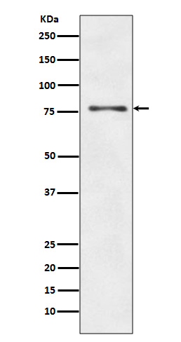
ATG7 Antibody
Rabbit mAb
- SPECIFICATION
- CITATIONS
- PROTOCOLS
- BACKGROUND

Application
| WB, IHC, FC, ICC, IP |
|---|---|
| Primary Accession | O95352 |
| Clonality | Monoclonal |
| Other Names | ATG7; APG7-LIKE; APG7L; DKFZp434N0735; GSA7; |
| Isotype | Rabbit IgG |
| Host | Rabbit |
| Calculated MW | 77960 Da |
| Dilution | WB 1:10000~1:50000 IHC 1:50~1:200 ICC/IF 1:50~1:200 IP 1:20 FC 1:20 |
|---|---|
| Purification | Affinity-chromatography |
| Immunogen | A synthesized peptide derived from human ATG7 |
| Description | The molecular machinery of autophagy was largely discovered in yeast and referred to as autophagy-related (Atg) genes. Formation of the autophagosome involves a ubiquitin-like conjugation system in which Atg12 is covalently bound to Atg5 and targeted to autophagosome vesicles. This conjugation reaction is mediated by the ubiquitin E1-like enzyme Atg7 and the E2-like enzyme Atg10. |
| Storage Condition and Buffer | Rabbit IgG in phosphate buffered saline , pH 7.4, 150mM NaCl, 0.02% sodium azide and 50% glycerol. Store at +4°C short term. Store at -20°C long term. Avoid freeze / thaw cycle. |
| Name | ATG7 (HGNC:16935) |
|---|---|
| Synonyms | APG7L |
| Function | E1-like activating enzyme involved in the 2 ubiquitin-like systems required for cytoplasm to vacuole transport (Cvt) and autophagy. Activates ATG12 for its conjugation with ATG5 as well as the ATG8 family proteins for their conjugation with phosphatidylethanolamine. Both systems are needed for the ATG8 association to Cvt vesicles and autophagosomes membranes. Required for autophagic death induced by caspase-8 inhibition. Facilitates LC3-I lipidation with phosphatidylethanolamine to form LC3-II which is found on autophagosomal membranes (PubMed:34161705). Required for mitophagy which contributes to regulate mitochondrial quantity and quality by eliminating the mitochondria to a basal level to fulfill cellular energy requirements and preventing excess ROS production. Modulates p53/TP53 activity to regulate cell cycle and survival during metabolic stress. Also plays a key role in the maintenance of axonal homeostasis, the prevention of axonal degeneration, the maintenance of hematopoietic stem cells, the formation of Paneth cell granules, as well as in adipose differentiation. Plays a role in regulating the liver clock and glucose metabolism by mediating the autophagic degradation of CRY1 (clock repressor) in a time-dependent manner (By similarity). |
| Cellular Location | Cytoplasm. Preautophagosomal structure. Note=Localizes also to discrete punctae along the ciliary axoneme and to the base of the ciliary axoneme |
| Tissue Location | Widely expressed, especially in kidney, liver, lymph nodes and bone marrow. |

Thousands of laboratories across the world have published research that depended on the performance of antibodies from Abcepta to advance their research. Check out links to articles that cite our products in major peer-reviewed journals, organized by research category.
info@abcepta.com, and receive a free "I Love Antibodies" mug.
Provided below are standard protocols that you may find useful for product applications.
If you have used an Abcepta product and would like to share how it has performed, please click on the "Submit Review" button and provide the requested information. Our staff will examine and post your review and contact you if needed.
If you have any additional inquiries please email technical services at tech@abcepta.com.


 Foundational characteristics of cancer include proliferation, angiogenesis, migration, evasion of apoptosis, and cellular immortality. Find key markers for these cellular processes and antibodies to detect them.
 The SUMOplot™ Analysis Program predicts and scores sumoylation sites in your protein. SUMOylation is a post-translational modification involved in various cellular processes, such as nuclear-cytosolic transport, transcriptional regulation, apoptosis, protein stability, response to stress, and progression through the cell cycle.
 The Autophagy Receptor Motif Plotter predicts and scores autophagy receptor binding sites in your protein. Identifying proteins connected to this pathway is critical to understanding the role of autophagy in physiological as well as pathological processes such as development, differentiation, neurodegenerative diseases, stress, infection, and cancer.


