


 Foundational characteristics of cancer include proliferation, angiogenesis, migration, evasion of apoptosis, and cellular immortality. Find key markers for these cellular processes and antibodies to detect them.
 The SUMOplot™ Analysis Program predicts and scores sumoylation sites in your protein. SUMOylation is a post-translational modification involved in various cellular processes, such as nuclear-cytosolic transport, transcriptional regulation, apoptosis, protein stability, response to stress, and progression through the cell cycle.
 The Autophagy Receptor Motif Plotter predicts and scores autophagy receptor binding sites in your protein. Identifying proteins connected to this pathway is critical to understanding the role of autophagy in physiological as well as pathological processes such as development, differentiation, neurodegenerative diseases, stress, infection, and cancer.
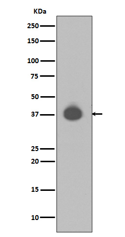
DUSP6 Antibody
Rabbit mAb
- SPECIFICATION
- CITATIONS
- PROTOCOLS
- BACKGROUND

Application
| WB, IHC, FC, ICC, IP |
|---|---|
| Primary Accession | Q16828 |
| Reactivity | Rat |
| Clonality | Monoclonal |
| Other Names | HH19; MKP3; PYST1; DUSP6; DUSP6a; Dual specificity phosphatase 6; |
| Isotype | Rabbit IgG |
| Host | Rabbit |
| Calculated MW | 42320 Da |
| Dilution | WB 1:500~1:2000 IHC 1:50~1:200 ICC/IF 1:50~1:200 IP 1:50 FC 1:50 |
|---|---|
| Purification | Affinity-chromatography |
| Immunogen | A synthesized peptide derived from human DUSP6 |
| Description | MAP kinases are inactivated by dual-specificity protein phosphatases (DUSP) that differ in their substrate specificity, tissue distribution, inducibility by extracellular stimuli and cellular localization. DUSPs, also known as MAPK phosphatases (MKP), specifically dephosphorylate both threonine and tyrosine residues in MAPK P-loops and have been shown to play important roles in regulating the function of the MAPK family. At least 13 members of the family (DUSP1-10, DUSp14, DUSP16, and DUSP22) display unique substrate specificities for various MAP kinases. |
| Storage Condition and Buffer | Rabbit IgG in phosphate buffered saline , pH 7.4, 150mM NaCl, 0.02% sodium azide and 50% glycerol. Store at +4°C short term. Store at -20°C long term. Avoid freeze / thaw cycle. |
| Name | DUSP6 |
|---|---|
| Synonyms | MKP3, PYST1 |
| Function | Inactivates MAP kinases. Has a specificity for the ERK family (PubMed:9858808). Plays an important role in alleviating chronic postoperative pain. Necessary for the normal dephosphorylation of the long-lasting phosphorylated forms of spinal MAPK1/3 and MAP kinase p38 induced by peripheral surgery, which drives the resolution of acute postoperative allodynia (By similarity). Also important for dephosphorylation of MAPK1/3 in local wound tissue, which further contributes to resolution of acute pain (By similarity). Promotes cell differentiation by regulating MAPK1/MAPK3 activity and regulating the expression of AP1 transcription factors (PubMed:29043977). |
| Cellular Location | Cytoplasm. |
| Tissue Location | Expressed in keratinocytes (at protein level). |
Research Areas
Citations (0)

Thousands of laboratories across the world have published research that depended on the performance of antibodies from Abcepta to advance their research. Check out links to articles that cite our products in major peer-reviewed journals, organized by research category.
Submit your citation using an Abcepta antibody to
info@abcepta.com, and receive a free "I Love Antibodies" mug.
info@abcepta.com, and receive a free "I Love Antibodies" mug.

Application Protocols
Provided below are standard protocols that you may find useful for product applications.
Abcepta welcomes feedback from its customers.
If you have used an Abcepta product and would like to share how it has performed, please click on the "Submit Review" button and provide the requested information. Our staff will examine and post your review and contact you if needed.
If you have any additional inquiries please email technical services at tech@abcepta.com.
$ 350.00
$ 175.00
Cat# AP90447
Ordering Information
United States
AlbaniaAustraliaAustriaBelgiumBosnia & HerzegovinaBrazilBulgariaCanadaCentral AmericaChinaCroatiaCyprusCzech RepublicDenmarkEstoniaFinlandFranceGermanyGreeceHong KongHungaryIcelandIndiaIndonesiaIrelandIsraelItalyJapanLatviaLithuaniaLuxembourgMacedoniaMalaysiaMaltaNetherlandsNew ZealandNorwayPakistanPolandPortugalRomaniaSerbiaSingaporeSlovakiaSloveniaSouth AfricaSouth KoreaSpainSwedenSwitzerlandTaiwanTurkeyUnited KingdomUnited StatesVietnamWorldwideOthersMexico
USA Headquarters
(888) 735-7227 / (858) 622-0099 or (858) 875-1900
Other Products
Shipping Information
Domestic orders (in stock items)
Shipped out the same day. Orders placed after 1 PM (PST) will ship out the next business day.
International orders
Contact your local distributors
