


 Foundational characteristics of cancer include proliferation, angiogenesis, migration, evasion of apoptosis, and cellular immortality. Find key markers for these cellular processes and antibodies to detect them.
 The SUMOplot™ Analysis Program predicts and scores sumoylation sites in your protein. SUMOylation is a post-translational modification involved in various cellular processes, such as nuclear-cytosolic transport, transcriptional regulation, apoptosis, protein stability, response to stress, and progression through the cell cycle.
 The Autophagy Receptor Motif Plotter predicts and scores autophagy receptor binding sites in your protein. Identifying proteins connected to this pathway is critical to understanding the role of autophagy in physiological as well as pathological processes such as development, differentiation, neurodegenerative diseases, stress, infection, and cancer.
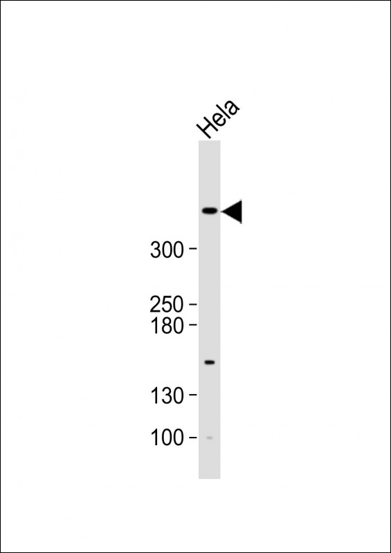
ATM Antibody (N-term)
Purified Rabbit Polyclonal Antibody (Pab)

- SPECIFICATION
- CITATIONS: 1
- PROTOCOLS
- BACKGROUND

Application
| IHC-P, WB, E |
|---|---|
| Primary Accession | Q13315 |
| Reactivity | Human |
| Host | Rabbit |
| Clonality | Polyclonal |
| Isotype | Rabbit IgG |
| Calculated MW | 350687 Da |
| Antigen Region | 5-34 aa |
| Gene ID | 472 |
|---|---|
| Other Names | Serine-protein kinase ATM, Ataxia telangiectasia mutated, A-T mutated, ATM |
| Target/Specificity | This ATM antibody is generated from rabbits immunized with a KLH conjugated synthetic peptide between 5~34 amino acids from the N-terminal region of human ATM. |
| Dilution | WB~~1:500 IHC-P~~1:50~100 |
| Format | Purified polyclonal antibody supplied in PBS with 0.09% (W/V) sodium azide. This antibody is prepared by Saturated Ammonium Sulfate (SAS) precipitation followed by dialysis against PBS. |
| Storage | Maintain refrigerated at 2-8°C for up to 2 weeks. For long term storage store at -20°C in small aliquots to prevent freeze-thaw cycles. |
| Precautions | ATM Antibody (N-term) is for research use only and not for use in diagnostic or therapeutic procedures. |
| Name | ATM |
|---|---|
| Function | Serine/threonine protein kinase which activates checkpoint signaling upon double strand breaks (DSBs), apoptosis and genotoxic stresses such as ionizing ultraviolet A light (UVA), thereby acting as a DNA damage sensor (PubMed:10550055, PubMed:10839545, PubMed:10910365, PubMed:12556884, PubMed:14871926, PubMed:15064416, PubMed:15448695, PubMed:15456891, PubMed:15790808, PubMed:15916964, PubMed:17923702, PubMed:21757780, PubMed:24534091, PubMed:35076389, PubMed:9733514). Recognizes the substrate consensus sequence [ST]-Q (PubMed:10550055, PubMed:10839545, PubMed:10910365, PubMed:12556884, PubMed:14871926, PubMed:15448695, PubMed:15456891, PubMed:15916964, PubMed:17923702, PubMed:24534091, PubMed:9733514). Phosphorylates 'Ser-139' of histone variant H2AX at double strand breaks (DSBs), thereby regulating DNA damage response mechanism (By similarity). Also plays a role in pre-B cell allelic exclusion, a process leading to expression of a single immunoglobulin heavy chain allele to enforce clonality and monospecific recognition by the B-cell antigen receptor (BCR) expressed on individual B-lymphocytes. After the introduction of DNA breaks by the RAG complex on one immunoglobulin allele, acts by mediating a repositioning of the second allele to pericentromeric heterochromatin, preventing accessibility to the RAG complex and recombination of the second allele. Also involved in signal transduction and cell cycle control. May function as a tumor suppressor. Necessary for activation of ABL1 and SAPK. Phosphorylates DYRK2, CHEK2, p53/TP53, FBXW7, FANCD2, NFKBIA, BRCA1, CREBBP/CBP, RBBP8/CTIP, MRE11, nibrin (NBN), RAD50, RAD17, PELI1, TERF1, UFL1, RAD9, UBQLN4 and DCLRE1C (PubMed:10550055, PubMed:10766245, PubMed:10802669, PubMed:10839545, PubMed:10910365, PubMed:10973490, PubMed:11375976, PubMed:12086603, PubMed:15456891, PubMed:19965871, PubMed:21757780, PubMed:24534091, PubMed:26240375, PubMed:26774286, PubMed:30612738, PubMed:30886146, PubMed:30952868, PubMed:38128537, PubMed:9733515, PubMed:9843217). May play a role in vesicle and/or protein transport. Could play a role in T-cell development, gonad and neurological function. Plays a role in replication-dependent histone mRNA degradation. Binds DNA ends. Phosphorylation of DYRK2 in nucleus in response to genotoxic stress prevents its MDM2-mediated ubiquitination and subsequent proteasome degradation (PubMed:19965871). Phosphorylates ATF2 which stimulates its function in DNA damage response (PubMed:15916964). Phosphorylates ERCC6 which is essential for its chromatin remodeling activity at DNA double- strand breaks (PubMed:29203878). Phosphorylates TTC5/STRAP at 'Ser-203' in the cytoplasm in response to DNA damage, which promotes TTC5/STRAP nuclear localization (PubMed:15448695). Also involved in pexophagy by mediating phosphorylation of PEX5: translocated to peroxisomes in response to reactive oxygen species (ROS), and catalyzes phosphorylation of PEX5, promoting PEX5 ubiquitination and induction of pexophagy (PubMed:26344566). |
| Cellular Location | Nucleus. Cytoplasmic vesicle. Cytoplasm, cytoskeleton, microtubule organizing center, centrosome {ECO:0000250|UniProtKB:Q62388}. Peroxisome matrix. Note=Primarily nuclear (PubMed:9050866, PubMed:9150358). Found also in endocytic vesicles in association with beta-adaptin (PubMed:9707615). Translocated to peroxisomes in response to reactive oxygen species (ROS) by PEX5 (PubMed:26344566) |
| Tissue Location | Found in pancreas, kidney, skeletal muscle, liver, lung, placenta, brain, heart, spleen, thymus, testis, ovary, small intestine, colon and leukocytes |

Provided below are standard protocols that you may find useful for product applications.
Background
ATM is involved in signal transduction, cell cycle control and DNA repair, and may function as a tumor suppressor. It is necessary for activation of ABL1 and SAPK, and phosphorylates p53, NFKBIA, BRCA1, CTIP, NIBRIN (NBS1), TERF1, and RAD9. This protein has potential roles in vesicle and/or protein transport, T-cell development, gonad and neurological function. ATM is also part of the BRCA1-associated genome surveillance complex. ATM is induced by ionizing radiation. Defects in ATM are the cause of ataxia talangiectasia (AT), also known as Louis-Bar syndrome, a rare recessive disorder characterized by progressive cerebellar ataxia, dilation of the blood vessels in the conjunctiva and eyeballs, immunodeficiency, growth retardation and sexual immaturity. About 30% of AT patients develop lymphomas and leukemias. Defects in ATM also contribute to T-cell acute lymphoblastic leukemia (TALL) and T-prolymphocytic leukemia (TPLL). TPLL is characterized by a high white blood cell count, with a predominance of prolymphocytes, marked splenomegaly, lymphadenopathy, skin lesions and serous effusion. Defects in ATM also contribute to B-cell non-Hodgkin's lymphomas, and to B-cell chronic lymphocytic leukemia, a disease characterized by accumulation of mature CD5+ B lymphocytes, lymphadenopathy, immunodeficiency and bone marrow failure.
References
Suzuki, A., et al., J. Biol. Chem. 278(1):48-53 (2003).
Kishi, S., et al., J. Biol. Chem. 276(31):29282-29291 (2001).
Schaffner, C., et al., Proc. Natl. Acad. Sci. U.S.A. 97(6):2773-2778 (2000).
Gatei, M., et al., Nat. Genet. 25(1):115-119 (2000).
Becker-Catania, S.G., et al., Mol. Genet. Metab. 70(2):122-133 (2000).
If you have used an Abcepta product and would like to share how it has performed, please click on the "Submit Review" button and provide the requested information. Our staff will examine and post your review and contact you if needed.
If you have any additional inquiries please email technical services at tech@abcepta.com.

Ordering Information
Other Products
Shipping Information
