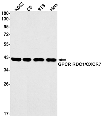
RDC1 Rabbit mAb
- SPECIFICATION
- CITATIONS
- PROTOCOLS
- BACKGROUND

Application
| WB |
|---|---|
| Primary Accession | P25106 |
| Reactivity | Human, Mouse, Rat |
| Host | Rabbit |
| Clonality | Monoclonal Antibody |
| Calculated MW | 41493 Da |
| Gene ID | 57007 |
|---|---|
| Other Names | ACKR3 |
| Dilution | WB~~1/500-1/1000 |
| Format | Liquid |
| Name | ACKR3 (HGNC:23692) |
|---|---|
| Function | Atypical chemokine receptor that controls chemokine levels and localization via high-affinity chemokine binding that is uncoupled from classic ligand-driven signal transduction cascades, resulting instead in chemokine sequestration, degradation, or transcytosis. Also known as interceptor (internalizing receptor) or chemokine-scavenging receptor or chemokine decoy receptor. Acts as a receptor for chemokines CXCL11 and CXCL12/SDF1 (PubMed:16107333, PubMed:19255243, PubMed:19380869, PubMed:20161793, PubMed:22300987). Chemokine binding does not activate G-protein-mediated signal transduction but instead induces beta-arrestin recruitment, leading to ligand internalization and activation of MAPK signaling pathway (PubMed:16940167, PubMed:18653785, PubMed:20018651). Required for regulation of CXCR4 protein levels in migrating interneurons, thereby adapting their chemokine responsiveness (PubMed:16940167, PubMed:18653785). In glioma cells, transduces signals via MEK/ERK pathway, mediating resistance to apoptosis. Promotes cell growth and survival (PubMed:16940167, PubMed:20388803). Not involved in cell migration, adhesion or proliferation of normal hematopoietic progenitors but activated by CXCL11 in malignant hemapoietic cells, leading to phosphorylation of ERK1/2 (MAPK3/MAPK1) and enhanced cell adhesion and migration (PubMed:17804806, PubMed:18653785, PubMed:19641136, PubMed:20887389). Plays a regulatory role in CXCR4-mediated activation of cell surface integrins by CXCL12 (PubMed:18653785). Required for heart valve development (PubMed:17804806). Regulates axon guidance in the oculomotor system through the regulation of CXCL12 levels (PubMed:31211835). |
| Cellular Location | Cell membrane; Multi-pass membrane protein. Early endosome. Recycling endosome. Note=Predominantly localizes to endocytic vesicles, and upon stimulation by the ligand is internalized via clathrin-coated pits in a beta-arrestin-dependent manner. Once internalized, the ligand dissociates from the receptor, and is targeted to degradation while the receptor is recycled back to the cell membrane. |
| Tissue Location | Expressed in monocytes, basophils, B-cells, umbilical vein endothelial cells (HUVEC) and B-lymphoblastoid cells Lower expression detected in CD4+ T-lymphocytes and natural killer cells. In the brain, detected in endothelial cells and capillaries, and in mature neurons of the frontal cortex and hippocampus. Expressed in tubular formation in the kidney. Highly expressed in astroglial tumor endothelial, microglial and glioma cells. Expressed at low levels in normal CD34+ progenitor cells, but at very high levels in several myeloid malignant cell lines. Expressed in breast carcinomas but not in normal breast tissue (at protein level). |

Thousands of laboratories across the world have published research that depended on the performance of antibodies from Abcepta to advance their research. Check out links to articles that cite our products in major peer-reviewed journals, organized by research category.
info@abcepta.com, and receive a free "I Love Antibodies" mug.
Provided below are standard protocols that you may find useful for product applications.
If you have used an Abcepta product and would like to share how it has performed, please click on the "Submit Review" button and provide the requested information. Our staff will examine and post your review and contact you if needed.
If you have any additional inquiries please email technical services at tech@abcepta.com.


 Foundational characteristics of cancer include proliferation, angiogenesis, migration, evasion of apoptosis, and cellular immortality. Find key markers for these cellular processes and antibodies to detect them.
 The SUMOplot™ Analysis Program predicts and scores sumoylation sites in your protein. SUMOylation is a post-translational modification involved in various cellular processes, such as nuclear-cytosolic transport, transcriptional regulation, apoptosis, protein stability, response to stress, and progression through the cell cycle.
 The Autophagy Receptor Motif Plotter predicts and scores autophagy receptor binding sites in your protein. Identifying proteins connected to this pathway is critical to understanding the role of autophagy in physiological as well as pathological processes such as development, differentiation, neurodegenerative diseases, stress, infection, and cancer.


