


 Foundational characteristics of cancer include proliferation, angiogenesis, migration, evasion of apoptosis, and cellular immortality. Find key markers for these cellular processes and antibodies to detect them.
 The SUMOplot™ Analysis Program predicts and scores sumoylation sites in your protein. SUMOylation is a post-translational modification involved in various cellular processes, such as nuclear-cytosolic transport, transcriptional regulation, apoptosis, protein stability, response to stress, and progression through the cell cycle.
 The Autophagy Receptor Motif Plotter predicts and scores autophagy receptor binding sites in your protein. Identifying proteins connected to this pathway is critical to understanding the role of autophagy in physiological as well as pathological processes such as development, differentiation, neurodegenerative diseases, stress, infection, and cancer.
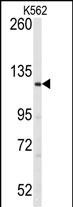
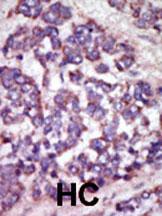
LATS1 Antibody (N-term)
Purified Rabbit Polyclonal Antibody (Pab)
- SPECIFICATION
- CITATIONS: 1
- PROTOCOLS
- BACKGROUND

Application
| IHC-P, WB, E |
|---|---|
| Primary Accession | O95835 |
| Reactivity | Human, Mouse, Rat |
| Host | Rabbit |
| Clonality | Polyclonal |
| Isotype | Rabbit IgG |
| Calculated MW | 126870 Da |
| Antigen Region | 1-30 aa |
| Gene ID | 9113 |
|---|---|
| Other Names | Serine/threonine-protein kinase LATS1, Large tumor suppressor homolog 1, WARTS protein kinase, h-warts, LATS1 {ECO:0000312|EMBL:AAD168821} |
| Target/Specificity | This LATS1 antibody is generated from rabbits immunized with a KLH conjugated synthetic peptide between 1-30 amino acids from the N-terminal region of human LATS1. |
| Dilution | WB~~1:1000 IHC-P~~1:50~100 |
| Format | Purified polyclonal antibody supplied in PBS with 0.09% (W/V) sodium azide. This antibody is prepared by Saturated Ammonium Sulfate (SAS) precipitation followed by dialysis against PBS. |
| Storage | Maintain refrigerated at 2-8°C for up to 2 weeks. For long term storage store at -20°C in small aliquots to prevent freeze-thaw cycles. |
| Precautions | LATS1 Antibody (N-term) is for research use only and not for use in diagnostic or therapeutic procedures. |
| Name | LATS1 {ECO:0000312|EMBL:AAD16882.1} |
|---|---|
| Function | Negative regulator of YAP1 in the Hippo signaling pathway that plays a pivotal role in organ size control and tumor suppression by restricting proliferation and promoting apoptosis (PubMed:10518011, PubMed:10831611, PubMed:18158288, PubMed:26437443, PubMed:28068668). The core of this pathway is composed of a kinase cascade wherein STK3/MST2 and STK4/MST1, in complex with its regulatory protein SAV1, phosphorylates and activates LATS1/2 in complex with its regulatory protein MOB1, which in turn phosphorylates and inactivates YAP1 oncoprotein and WWTR1/TAZ (PubMed:18158288, PubMed:26437443, PubMed:28068668). Phosphorylation of YAP1 by LATS1 inhibits its translocation into the nucleus to regulate cellular genes important for cell proliferation, cell death, and cell migration (PubMed:18158288, PubMed:26437443, PubMed:28068668). Acts as a tumor suppressor which plays a critical role in maintenance of ploidy through its actions in both mitotic progression and the G1 tetraploidy checkpoint (PubMed:15122335, PubMed:19927127). Negatively regulates G2/M transition by down-regulating CDK1 kinase activity (PubMed:9988268). Involved in the control of p53 expression (PubMed:15122335). Affects cytokinesis by regulating actin polymerization through negative modulation of LIMK1 (PubMed:15220930). May also play a role in endocrine function. Plays a role in mammary gland epithelial cell differentiation, both through the Hippo signaling pathway and the intracellular estrogen receptor signaling pathway by promoting the degradation of ESR1 (PubMed:28068668). Acts as an activator of the NLRP3 inflammasome by mediating phosphorylation of 'Ser-265' of NLRP3 following NLRP3 palmitoylation, promoting NLRP3 activation by NEK7 (PubMed:39173637). |
| Cellular Location | Cytoplasm, cytoskeleton, microtubule organizing center, centrosome. Cytoplasm, cytoskeleton, spindle. Midbody. Cytoplasm, cytoskeleton, microtubule organizing center, spindle pole body Note=Localizes to the centrosomes throughout interphase but migrates to the mitotic apparatus, including spindle pole bodies, mitotic spindle, and midbody, during mitosis. |
| Tissue Location | Expressed in all adult tissues examined except for lung and kidney. |

Provided below are standard protocols that you may find useful for product applications.
Background
The protein encoded by this gene is a putative serine/threonine kinase that localizes to the mitotic apparatus and complexes with cell cycle controller CDC2 kinase in early mitosis. The protein is phosphorylated in a cell-cycle dependent manner, with late prophase phosphorylation remaining through metaphase. The N-terminal region of the protein binds CDC2 to form a complex showing reduced H1 histone kinase activity, indicating a role as a negative regulator of CDC2/cyclin A. In addition, the C-terminal kinase domain binds to its own N-terminal region, suggesting potential negative regulation through interference with complex formation via intramolecular binding. Biochemical and genetic data suggest a role as a tumor suppressor. This is supported by studies in knockout mice showing development of soft-tissue sarcomas, ovarian stromal cell tumors and a high sensitivity to carcinogenic treatments.
References
Iida, S., et al., Oncogene 23(31):5266-5274 (2004).
Yang, X., et al., Nat. Cell Biol. 6(7):609-617 (2004).
Kamikubo, Y., et al., J. Biol. Chem. 278(20):17609-17614 (2003).
Hisaoka, M., et al., Lab. Invest. 82(10):1427-1435 (2002).
Hirota, T., et al., J. Cell Biol. 149(5):1073-1086 (2000).
If you have used an Abcepta product and would like to share how it has performed, please click on the "Submit Review" button and provide the requested information. Our staff will examine and post your review and contact you if needed.
If you have any additional inquiries please email technical services at tech@abcepta.com.

Ordering Information
Other Products
Shipping Information
