


 Foundational characteristics of cancer include proliferation, angiogenesis, migration, evasion of apoptosis, and cellular immortality. Find key markers for these cellular processes and antibodies to detect them.
 The SUMOplot™ Analysis Program predicts and scores sumoylation sites in your protein. SUMOylation is a post-translational modification involved in various cellular processes, such as nuclear-cytosolic transport, transcriptional regulation, apoptosis, protein stability, response to stress, and progression through the cell cycle.
 The Autophagy Receptor Motif Plotter predicts and scores autophagy receptor binding sites in your protein. Identifying proteins connected to this pathway is critical to understanding the role of autophagy in physiological as well as pathological processes such as development, differentiation, neurodegenerative diseases, stress, infection, and cancer.
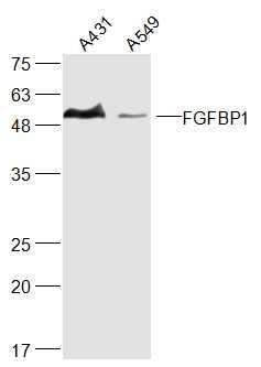
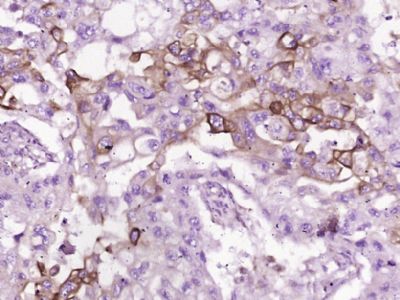
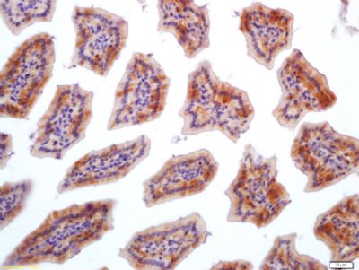
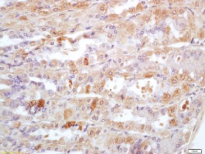
FGFBP1 Polyclonal Antibody
Purified Rabbit Polyclonal Antibody (Pab)
- SPECIFICATION
- CITATIONS
- PROTOCOLS
- BACKGROUND

Application
| WB, IHC-P, IHC-F, IF, E |
|---|---|
| Primary Accession | Q14512 |
| Reactivity | Rat |
| Host | Rabbit |
| Clonality | Polyclonal |
| Calculated MW | 26264 Da |
| Gene ID | 9982 |
|---|---|
| Other Names | Fibroblast growth factor-binding protein 1, FGF-BP, FGF-BP1, FGF-binding protein 1, FGFBP-1, 17 kDa heparin-binding growth factor-binding protein, 17 kDa HBGF-binding protein, HBp17, FGFBP1, FGFBP, HBP17 |
| Dilution | WB=1:500-2000,IHC-P=1:100-500,IHC-F=1:100-500,IF=1:100-500,ELISA=1:5000-10000 |
| Format | 0.01M TBS(pH7.4), 0.09% (W/V) sodium azide and 50% Glyce |
| Storage | Store at -20 ℃ for one year. Avoid repeated freeze/thaw cycles. When reconstituted in sterile pH 7.4 0.01M PBS or diluent of antibody the antibody is stable for at least two weeks at 2-4 ℃. |
| Name | FGFBP1 |
|---|---|
| Synonyms | FGFBP, HBP17 |
| Function | Acts as a carrier protein that release fibroblast-binding factors (FGFs) from the extracellular matrix (EM) storage and thus enhance the mitogenic activity of FGFs. Enhances FGF2 signaling during tissue repair, angiogenesis and in tumor growth. |
| Cellular Location | Secreted, extracellular space. Cell membrane; Peripheral membrane protein. Note=Extracellular and plasma membrane- associated. Colocalizes with HSPG2 in the pericellular environment of squamous cell carcinomas. |
| Tissue Location | Expressed in the suprabasal region of the epidermis, in hair follicles, the basement membrane at the dermo- epidermal junction (occasionally extending into the basement membrane of dermal blood vessels), wounded skin and several invasive squamous cell carcinomas (at protein level). Expressed in normal and wounded skin and various squamous cell carcinomas |
Research Areas
Citations (0)

Thousands of laboratories across the world have published research that depended on the performance of antibodies from Abcepta to advance their research. Check out links to articles that cite our products in major peer-reviewed journals, organized by research category.
Submit your citation using an Abcepta antibody to
info@abcepta.com, and receive a free "I Love Antibodies" mug.
info@abcepta.com, and receive a free "I Love Antibodies" mug.

Application Protocols
Provided below are standard protocols that you may find useful for product applications.
Abcepta welcomes feedback from its customers.
If you have used an Abcepta product and would like to share how it has performed, please click on the "Submit Review" button and provide the requested information. Our staff will examine and post your review and contact you if needed.
If you have any additional inquiries please email technical services at tech@abcepta.com.
$ 350.00
Cat# AP56797
Ordering Information
United States
AlbaniaAustraliaAustriaBelgiumBosnia & HerzegovinaBrazilBulgariaCanadaCentral AmericaChinaCroatiaCyprusCzech RepublicDenmarkEstoniaFinlandFranceGermanyGreeceHong KongHungaryIcelandIndiaIndonesiaIrelandIsraelItalyJapanLatviaLithuaniaLuxembourgMacedoniaMalaysiaMaltaNetherlandsNew ZealandNorwayPakistanPolandPortugalRomaniaSerbiaSingaporeSlovakiaSloveniaSouth AfricaSouth KoreaSpainSwedenSwitzerlandTaiwanTurkeyUnited KingdomUnited StatesVietnamWorldwideOthers
USA Headquarters
(888) 735-7227 / (858) 622-0099 or (858) 875-1900
Other Products
Shipping Information
Domestic orders (in stock items)
Shipped out the same day. Orders placed after 1 PM (PST) will ship out the next business day.
International orders
Contact your local distributors
