


 Foundational characteristics of cancer include proliferation, angiogenesis, migration, evasion of apoptosis, and cellular immortality. Find key markers for these cellular processes and antibodies to detect them.
 The SUMOplot™ Analysis Program predicts and scores sumoylation sites in your protein. SUMOylation is a post-translational modification involved in various cellular processes, such as nuclear-cytosolic transport, transcriptional regulation, apoptosis, protein stability, response to stress, and progression through the cell cycle.
 The Autophagy Receptor Motif Plotter predicts and scores autophagy receptor binding sites in your protein. Identifying proteins connected to this pathway is critical to understanding the role of autophagy in physiological as well as pathological processes such as development, differentiation, neurodegenerative diseases, stress, infection, and cancer.
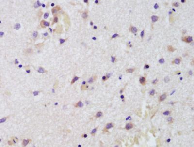
FLRT3 Polyclonal Antibody
Purified Rabbit Polyclonal Antibody (Pab)
- SPECIFICATION
- CITATIONS
- PROTOCOLS
- BACKGROUND

Application
| WB, E |
|---|---|
| Primary Accession | Q9NZU0 |
| Reactivity | Rat, Pig, Dog, Bovine |
| Host | Rabbit |
| Clonality | Polyclonal |
| Calculated MW | 73004 Da |
| Gene ID | 23767 |
|---|---|
| Other Names | Leucine-rich repeat transmembrane protein FLRT3, Fibronectin-like domain-containing leucine-rich transmembrane protein 3, FLRT3, KIAA1469 |
| Dilution | WB=1:500-2000,ELISA=1:5000-10000 |
| Format | 0.01M TBS(pH7.4), 0.09% (W/V) sodium azide and 50% Glyce |
| Storage | Store at -20 ℃ for one year. Avoid repeated freeze/thaw cycles. When reconstituted in sterile pH 7.4 0.01M PBS or diluent of antibody the antibody is stable for at least two weeks at 2-4 ℃. |
| Name | FLRT3 |
|---|---|
| Synonyms | KIAA1469 |
| Function | Functions in cell-cell adhesion, cell migration and axon guidance, exerting an attractive or repulsive role depending on its interaction partners. Plays a role in the spatial organization of brain neurons. Plays a role in vascular development in the retina (By similarity). Plays a role in cell-cell adhesion via its interaction with ADGRL3 and probably also other latrophilins that are expressed at the surface of adjacent cells (PubMed:26235030). Interaction with the intracellular domain of ROBO1 mediates axon attraction towards cells expressing NTN1. Mediates axon growth cone collapse and plays a repulsive role in neuron guidance via its interaction with UNC5B, and possibly also other UNC-5 family members (By similarity). Promotes neurite outgrowth (in vitro) (PubMed:14706654). Mediates cell-cell contacts that promote an increase both in neurite number and in neurite length. Plays a role in the regulation of the density of glutamaergic synapses. Plays a role in fibroblast growth factor-mediated signaling cascades. Required for normal morphogenesis during embryonic development, but not for normal embryonic patterning. Required for normal ventral closure, headfold fusion and definitive endoderm migration during embryonic development. Required for the formation of a normal basement membrane and the maintenance of a normal anterior visceral endoderm during embryonic development (By similarity). |
| Cellular Location | Cell membrane {ECO:0000250|UniProtKB:Q8BGT1}; Single-pass membrane protein {ECO:0000250|UniProtKB:Q8BGT1} Endoplasmic reticulum membrane {ECO:0000250|UniProtKB:Q8BGT1}. Cell junction, focal adhesion {ECO:0000250|UniProtKB:Q8BGT1}. Secreted {ECO:0000250|UniProtKB:Q8BGT1}. Cell projection, axon {ECO:0000250|UniProtKB:Q8BGT1}. Cell projection, growth cone membrane {ECO:0000250|UniProtKB:Q8BGT1}. Note=Detected on dendritic punctae that colocalize in part with glutamaergic synapses, but not with GABAergic synapses. Proteolytic cleavage in the juxtamembrane region gives rise to a shedded ectodomain. {ECO:0000250|UniProtKB:B1H234, ECO:0000250|UniProtKB:Q8BGT1} |
| Tissue Location | Expressed in kidney, brain, pancreas, skeletal muscle, lung, liver, placenta, and heart |
Research Areas
Citations (0)

Thousands of laboratories across the world have published research that depended on the performance of antibodies from Abcepta to advance their research. Check out links to articles that cite our products in major peer-reviewed journals, organized by research category.
Submit your citation using an Abcepta antibody to
info@abcepta.com, and receive a free "I Love Antibodies" mug.
info@abcepta.com, and receive a free "I Love Antibodies" mug.

Application Protocols
Provided below are standard protocols that you may find useful for product applications.
Abcepta welcomes feedback from its customers.
If you have used an Abcepta product and would like to share how it has performed, please click on the "Submit Review" button and provide the requested information. Our staff will examine and post your review and contact you if needed.
If you have any additional inquiries please email technical services at tech@abcepta.com.
$ 350.00
Cat# AP54837
Ordering Information
United States
AlbaniaAustraliaAustriaBelgiumBosnia & HerzegovinaBrazilBulgariaCanadaCentral AmericaChinaCroatiaCyprusCzech RepublicDenmarkEstoniaFinlandFranceGermanyGreeceHong KongHungaryIcelandIndiaIndonesiaIrelandIsraelItalyJapanLatviaLithuaniaLuxembourgMacedoniaMalaysiaMaltaNetherlandsNew ZealandNorwayPakistanPolandPortugalRomaniaSerbiaSingaporeSlovakiaSloveniaSouth AfricaSouth KoreaSpainSwedenSwitzerlandTaiwanTurkeyUnited KingdomUnited StatesVietnamWorldwideOthers
USA Headquarters
(888) 735-7227 / (858) 622-0099 or (858) 875-1900
Other Products
Shipping Information
Domestic orders (in stock items)
Shipped out the same day. Orders placed after 1 PM (PST) will ship out the next business day.
International orders
Contact your local distributors
