


 Foundational characteristics of cancer include proliferation, angiogenesis, migration, evasion of apoptosis, and cellular immortality. Find key markers for these cellular processes and antibodies to detect them.
 The SUMOplot™ Analysis Program predicts and scores sumoylation sites in your protein. SUMOylation is a post-translational modification involved in various cellular processes, such as nuclear-cytosolic transport, transcriptional regulation, apoptosis, protein stability, response to stress, and progression through the cell cycle.
 The Autophagy Receptor Motif Plotter predicts and scores autophagy receptor binding sites in your protein. Identifying proteins connected to this pathway is critical to understanding the role of autophagy in physiological as well as pathological processes such as development, differentiation, neurodegenerative diseases, stress, infection, and cancer.
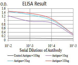
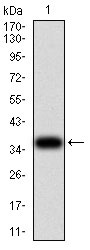
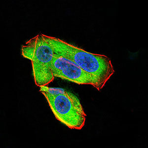
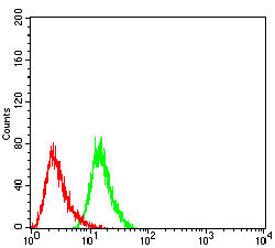
UCP2 Antibody
Purified Mouse Monoclonal Antibody

- SPECIFICATION
- CITATIONS
- PROTOCOLS
- BACKGROUND

Application
| WB, FC, ICC, E |
|---|---|
| Primary Accession | P55851 |
| Reactivity | Human |
| Host | Mouse |
| Clonality | Monoclonal |
| Clone Names | 3F1B9 |
| Isotype | IgG2a |
| Calculated MW | 33.2kDa |
| Description | Mitochondrial uncoupling proteins (UCP) are members of the larger family of mitochondrial anion carrier proteins (MACP). UCPs separate oxidative phosphorylation from ATP synthesis with energy dissipated as heat, also referred to as the mitochondrial proton leak. UCPs facilitate the transfer of anions from the inner to the outer mitochondrial membrane and the return transfer of protons from the outer to the inner mitochondrial membrane. They also reduce the mitochondrial membrane potential in mammalian cells. Tissue specificity occurs for the different UCPs and the exact methods of how UCPs transfer H+/OH- are not known. UCPs contain the three homologous protein domains of MACPs. This gene is expressed in many tissues, with the greatest expression in skeletal muscle. It is thought to play a role in nonshivering thermogenesis, obesity and diabetes. Chromosomal order is 5'-UCP3-UCP2-3'. |
| Immunogen | Purified recombinant fragment of human UCP2 (AA: 1-309) expressed in E. Coli. |
| Formulation | Purified antibody in PBS with 0.05% sodium azide |
| Gene ID | 7351 |
|---|---|
| Other Names | Mitochondrial uncoupling protein 2, UCP 2, Solute carrier family 25 member 8, UCPH, UCP2, SLC25A8 |
| Dilution | E~~1/10000 WB~~1/500 - 1/2000 IF~~1/200 - 1/1000 FC~~1/200 - 1/400 |
| Storage | Maintain refrigerated at 2-8°C for up to 6 months. For long term storage store at -20°C in small aliquots to prevent freeze-thaw cycles. |
| Precautions | UCP2 Antibody is for research use only and not for use in diagnostic or therapeutic procedures. |
| Name | UCP2 |
|---|---|
| Synonyms | SLC25A8 {ECO:0000303|PubMed:33798544} |
| Function | Antiporter that exports dicarboxylate intermediates of the Krebs cycle in exchange for phosphate plus a proton across the inner membrane of mitochondria, a process driven by mitochondrial motive force with an overall impact on glycolysis, glutaminolysis and glutathione-dependent redox balance. Continuous export of oxaloacetate and related four-carbon dicarboxylates from mitochondrial matrix into the cytosol negatively regulates the oxidation of acetyl-CoA substrates via the Krebs cycle, lowering the ATP/ADP ratio and reactive oxygen species (ROS) production (PubMed:24395786). May mediate inducible proton entry into the mitochondrial matrix affecting ATP turnover as a protection mechanism against oxidative stress. The proton currents are most likely associated with fatty acid flipping across the inner membrane of mitochondria in a metabolic process regulated by free fatty acids and purine nucleotides (PubMed:11171965, PubMed:33373220, PubMed:11278935, PubMed:22524567, PubMed:26182433) (By similarity). Regulates the use of glucose as a source of energy. Required for glucose-induced DRP1-dependent mitochondrial fission and neuron activation in the ventromedial nucleus of the hypothalamus (VMH). This mitochondrial adaptation mechanism modulates the VMH pool of glucose- excited neurons with an impact on systemic glucose homeostasis (By similarity). Regulates ROS levels and metabolic reprogramming of macrophages during the resolution phase of inflammation. Attenuates ROS production in response to IL33 to preserve the integrity of the Krebs cycle required for persistent production of itaconate and subsequent GATA3-dependent differentiation of inflammation-resolving alternatively activated macrophages (By similarity). Can unidirectionally transport anions including L-malate, L-aspartate, phosphate and chloride ions (PubMed:24395786, PubMed:22524567, PubMed:26182433). Does not mediate adaptive thermogenesis (By similarity). |
| Cellular Location | Mitochondrion inner membrane {ECO:0000250|UniProtKB:P70406}; Multi-pass membrane protein |
| Tissue Location | Widely expressed in adult human tissues, including tissues rich in macrophages. Most expressed in white adipose tissue and skeletal muscle. |

Thousands of laboratories across the world have published research that depended on the performance of antibodies from Abcepta to advance their research. Check out links to articles that cite our products in major peer-reviewed journals, organized by research category.
info@abcepta.com, and receive a free "I Love Antibodies" mug.

Provided below are standard protocols that you may find useful for product applications.
References
1.Endocrine. 2013 Jun;43(3):714-23. 2.Carcinogenesis. 2012 Nov;33(11):2065-75.
If you have used an Abcepta product and would like to share how it has performed, please click on the "Submit Review" button and provide the requested information. Our staff will examine and post your review and contact you if needed.
If you have any additional inquiries please email technical services at tech@abcepta.com.
Ordering Information
Other Products
Shipping Information
