


 Foundational characteristics of cancer include proliferation, angiogenesis, migration, evasion of apoptosis, and cellular immortality. Find key markers for these cellular processes and antibodies to detect them.
 The SUMOplot™ Analysis Program predicts and scores sumoylation sites in your protein. SUMOylation is a post-translational modification involved in various cellular processes, such as nuclear-cytosolic transport, transcriptional regulation, apoptosis, protein stability, response to stress, and progression through the cell cycle.
 The Autophagy Receptor Motif Plotter predicts and scores autophagy receptor binding sites in your protein. Identifying proteins connected to this pathway is critical to understanding the role of autophagy in physiological as well as pathological processes such as development, differentiation, neurodegenerative diseases, stress, infection, and cancer.
DAPK3 Antibody
Purified Mouse Monoclonal Antibody
- SPECIFICATION
- CITATIONS
- PROTOCOLS
- BACKGROUND

Application
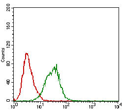
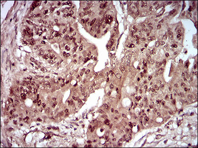
| WB, IHC, FC, E |
|---|---|
| Primary Accession | O43293 |
| Reactivity | Human |
| Host | Mouse |
| Clonality | Monoclonal |
| Clone Names | 4H4C8 |
| Isotype | IgG1 |
| Calculated MW | 52.5kDa |
| Description | Death-associated protein kinase 3 (DAPK3) induces morphological changes in apoptosis when overexpressed in mammalian cells. These results suggest that DAPK3 may play a role in the induction of apoptosis. |
| Immunogen | Purified recombinant fragment of human DAPK3 (AA: 28-161) expressed in E. Coli. |
| Formulation | Purified antibody in PBS with 0.05% sodium azide. |
| Gene ID | 1613 |
|---|---|
| Other Names | Death-associated protein kinase 3, DAP kinase 3, 2.7.11.1, DAP-like kinase, Dlk, MYPT1 kinase, Zipper-interacting protein kinase, ZIP-kinase, DAPK3, ZIPK |
| Dilution | E~~1/10000 WB~~1/500 - 1/2000 FC~~1/200 - 1/400 IHC~~1/200 - 1/1000 |
| Storage | Maintain refrigerated at 2-8°C for up to 6 months. For long term storage store at -20°C in small aliquots to prevent freeze-thaw cycles. |
| Precautions | DAPK3 Antibody is for research use only and not for use in diagnostic or therapeutic procedures. |
| Name | DAPK3 |
|---|---|
| Synonyms | ZIPK |
| Function | Serine/threonine kinase which is involved in the regulation of apoptosis, autophagy, transcription, translation and actin cytoskeleton reorganization. Involved in the regulation of smooth muscle contraction. Regulates both type I (caspase-dependent) apoptotic and type II (caspase-independent) autophagic cell deaths signal, depending on the cellular setting. Involved in regulation of starvation-induced autophagy. Regulates myosin phosphorylation in both smooth muscle and non-muscle cells. In smooth muscle, regulates myosin either directly by phosphorylating MYL12B and MYL9 or through inhibition of smooth muscle myosin phosphatase (SMPP1M) via phosphorylation of PPP1R12A; the inhibition of SMPP1M functions to enhance muscle responsiveness to Ca(2+) and promote a contractile state. Phosphorylates MYL12B in non-muscle cells leading to reorganization of actin cytoskeleton. Isoform 2 can phosphorylate myosin, PPP1R12A and MYL12B. Overexpression leads to condensation of actin stress fibers into thick bundles. Involved in actin filament focal adhesion dynamics. The function in both reorganization of actin cytoskeleton and focal adhesion dissolution is modulated by RhoD. Positively regulates canonical Wnt/beta-catenin signaling through interaction with NLK and TCF7L2. Phosphorylates RPL13A on 'Ser-77' upon interferon-gamma activation which is causing RPL13A release from the ribosome, RPL13A association with the GAIT complex and its subsequent involvement in transcript-selective translation inhibition. Enhances transcription from AR-responsive promoters in a hormone- and kinase- dependent manner. Involved in regulation of cell cycle progression and cell proliferation. May be a tumor suppressor. |
| Cellular Location | Nucleus. Nucleus, PML body {ECO:0000250|UniProtKB:O54784}. Cytoplasm, cytoskeleton, microtubule organizing center, centrosome {ECO:0000250|UniProtKB:O54784}. Chromosome, centromere. Cytoplasm. Cytoplasm, cytoskeleton, spindle. Midbody Note=Predominantly localizes to the cytoplasm but can shuttle between the nucleus and cytoplasm; cytoplasmic localization is promoted by phosphorylation at Thr-299 and involves Rho/Rock signaling [Isoform 2]: Nucleus. Cytoplasm |
| Tissue Location | Widely expressed. Isoform 1 and isoform 2 are expressed in the bladder smooth muscle. |

Thousands of laboratories across the world have published research that depended on the performance of antibodies from Abcepta to advance their research. Check out links to articles that cite our products in major peer-reviewed journals, organized by research category.
info@abcepta.com, and receive a free "I Love Antibodies" mug.

Provided below are standard protocols that you may find useful for product applications.
Background
The protein encoded by this gene is an isozyme of very long-chain acyl-CoA synthetase (VLCS). It is capable of activating very long-chain fatty-acids containing 24- and 26-carbons. It is expressed in liver and associated with endoplasmic reticulum but not with peroxisomes. Its primary role is in fatty acid elongation or complex lipid synthesis rather than in degradation. This gene has a mouse ortholog. ; ;
References
1. Cancer Res. 2011 Apr 15;71(8):3152-61.2. Int J Cancer. 2009 Apr 1;124(7):1587-93.
If you have used an Abcepta product and would like to share how it has performed, please click on the "Submit Review" button and provide the requested information. Our staff will examine and post your review and contact you if needed.
If you have any additional inquiries please email technical services at tech@abcepta.com.
Ordering Information
Other Products
Shipping Information
