


 Foundational characteristics of cancer include proliferation, angiogenesis, migration, evasion of apoptosis, and cellular immortality. Find key markers for these cellular processes and antibodies to detect them.
 The SUMOplot™ Analysis Program predicts and scores sumoylation sites in your protein. SUMOylation is a post-translational modification involved in various cellular processes, such as nuclear-cytosolic transport, transcriptional regulation, apoptosis, protein stability, response to stress, and progression through the cell cycle.
 The Autophagy Receptor Motif Plotter predicts and scores autophagy receptor binding sites in your protein. Identifying proteins connected to this pathway is critical to understanding the role of autophagy in physiological as well as pathological processes such as development, differentiation, neurodegenerative diseases, stress, infection, and cancer.
> home > Products > Primary Antibodies > Antibody Collections > Jak-STAT signaling KEGG > STAT1 Antibody
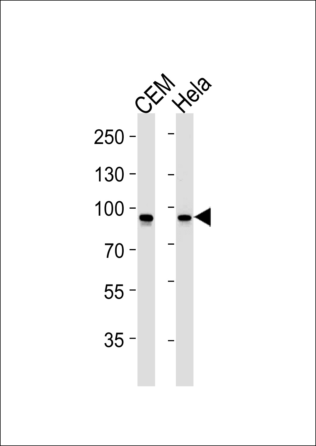
STAT1 Antibody
Mouse Monoclonal Antibody (Mab)
- SPECIFICATION
- CITATIONS
- PROTOCOLS
- BACKGROUND

Application
| WB, E |
|---|---|
| Primary Accession | P42224 |
| Reactivity | Human |
| Host | Mouse |
| Clonality | Monoclonal |
| Isotype | IgG1 |
| Clone/Animal Names | 1141CT26.2.1 |
| Calculated MW | 87335 Da |
| Gene ID | 6772 |
|---|---|
| Other Names | Signal transducer and activator of transcription 1-alpha/beta, Transcription factor ISGF-3 components p91/p84, STAT1 |
| Target/Specificity | Purified His-tagged STAT1 protein was used to produced this monoclonal antibody. |
| Dilution | WB~~1:1000 |
| Format | Purified monoclonal antibody supplied in PBS with 0.09% (W/V) sodium azide. This antibody is purified through a protein G column, followed by dialysis against PBS. |
| Storage | Maintain refrigerated at 2-8°C for up to 2 weeks. For long term storage store at -20°C in small aliquots to prevent freeze-thaw cycles. |
| Precautions | STAT1 Antibody is for research use only and not for use in diagnostic or therapeutic procedures. |
| Name | STAT1 |
|---|---|
| Function | Signal transducer and transcription activator that mediates cellular responses to interferons (IFNs), cytokine KITLG/SCF and other cytokines and other growth factors (PubMed:12764129, PubMed:12855578, PubMed:15322115, PubMed:23940278, PubMed:34508746, PubMed:35568036, PubMed:9724754). Following type I IFN (IFN-alpha and IFN-beta) binding to cell surface receptors, signaling via protein kinases leads to activation of Jak kinases (TYK2 and JAK1) and to tyrosine phosphorylation of STAT1 and STAT2. The phosphorylated STATs dimerize and associate with ISGF3G/IRF-9 to form a complex termed ISGF3 transcription factor, that enters the nucleus (PubMed:28753426, PubMed:35568036). ISGF3 binds to the IFN stimulated response element (ISRE) to activate the transcription of IFN-stimulated genes (ISG), which drive the cell in an antiviral state (PubMed:28753426, PubMed:35568036). In response to type II IFN (IFN-gamma), STAT1 is tyrosine- and serine-phosphorylated (PubMed:26479788). It then forms a homodimer termed IFN-gamma-activated factor (GAF), migrates into the nucleus and binds to the IFN gamma activated sequence (GAS) to drive the expression of the target genes, inducing a cellular antiviral state (PubMed:8156998). Becomes activated in response to KITLG/SCF and KIT signaling (PubMed:15526160). May mediate cellular responses to activated FGFR1, FGFR2, FGFR3 and FGFR4 (PubMed:19088846). Following bacterial lipopolysaccharide (LPS)-induced TLR4 endocytosis, phosphorylated at Thr-749 by IKBKB which promotes binding of STAT1 to the 5'-TTTGAGGC-3' sequence in the ARID5A promoter, resulting in transcriptional activation of ARID5A and subsequent ARID5A-mediated stabilization of IL6 (PubMed:32209697). Phosphorylation at Thr-749 also promotes binding of STAT1 to the 5'-TTTGAGTC-3' sequence in the IL12B promoter and activation of IL12B transcription (PubMed:32209697). Involved in food tolerance in small intestine: associates with the Gasdermin-D, p13 cleavage product (13 kDa GSDMD) and promotes transcription of CIITA, inducing type 1 regulatory T (Tr1) cells in upper small intestine (By similarity). |
| Cellular Location | Cytoplasm. Nucleus Note=Translocated into the nucleus upon tyrosine phosphorylation and dimerization, in response to IFN-gamma and signaling by activated FGFR1, FGFR2, FGFR3 or FGFR4 (PubMed:15322115). Monomethylation at Lys- 525 is required for phosphorylation at Tyr-701 and translocation into the nucleus (PubMed:28753426). Translocates into the nucleus in response to interferon-beta stimulation (PubMed:26479788) |
Research Areas

Abcepta welcomes feedback from its customers.
If you have used an Abcepta product and would like to share how it has performed, please click on the "Submit Review" button and provide the requested information. Our staff will examine and post your review and contact you if needed.
If you have any additional inquiries please email technical services at tech@abcepta.com.
$ 385.00
Cat# AM2229b
Ordering Information
United States
AlbaniaAustraliaAustriaBelgiumBosnia & HerzegovinaBrazilBulgariaCanadaCentral AmericaChinaCroatiaCyprusCzech RepublicDenmarkEstoniaFinlandFranceGermanyGreeceHong KongHungaryIcelandIndiaIndonesiaIrelandIsraelItalyJapanLatviaLithuaniaLuxembourgMacedoniaMalaysiaMaltaNetherlandsNew ZealandNorwayPakistanPolandPortugalRomaniaSerbiaSingaporeSlovakiaSloveniaSouth AfricaSouth KoreaSpainSwedenSwitzerlandTaiwanTurkeyUnited KingdomUnited StatesVietnamWorldwideOthersMexico
USA Headquarters
(888) 735-7227 / (858) 622-0099 or (858) 875-1900
Other Products
Shipping Information
Domestic orders (in stock items)
Shipped out the same day. Orders placed after 1 PM (PST) will ship out the next business day.
International orders
Contact your local distributors
