


 Foundational characteristics of cancer include proliferation, angiogenesis, migration, evasion of apoptosis, and cellular immortality. Find key markers for these cellular processes and antibodies to detect them.
 The SUMOplot™ Analysis Program predicts and scores sumoylation sites in your protein. SUMOylation is a post-translational modification involved in various cellular processes, such as nuclear-cytosolic transport, transcriptional regulation, apoptosis, protein stability, response to stress, and progression through the cell cycle.
 The Autophagy Receptor Motif Plotter predicts and scores autophagy receptor binding sites in your protein. Identifying proteins connected to this pathway is critical to understanding the role of autophagy in physiological as well as pathological processes such as development, differentiation, neurodegenerative diseases, stress, infection, and cancer.
Anti-RAGE/AGER Antibody Picoband™ (monoclonal, 5C6C1)
- SPECIFICATION
- CITATIONS
- PROTOCOLS
- BACKGROUND

Application
| WB, IHC, FC |
|---|---|
| Primary Accession | Q15109 |
| Host | Mouse |
| Isotype | IgG2b |
| Reactivity | Rat, Human, Mouse |
| Clonality | Monoclonal |
| Format | Lyophilized |
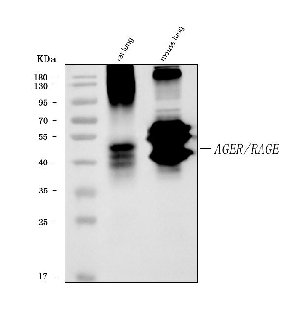
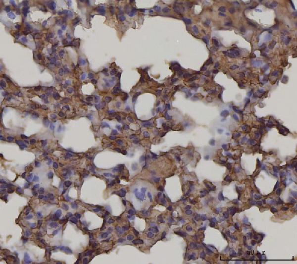
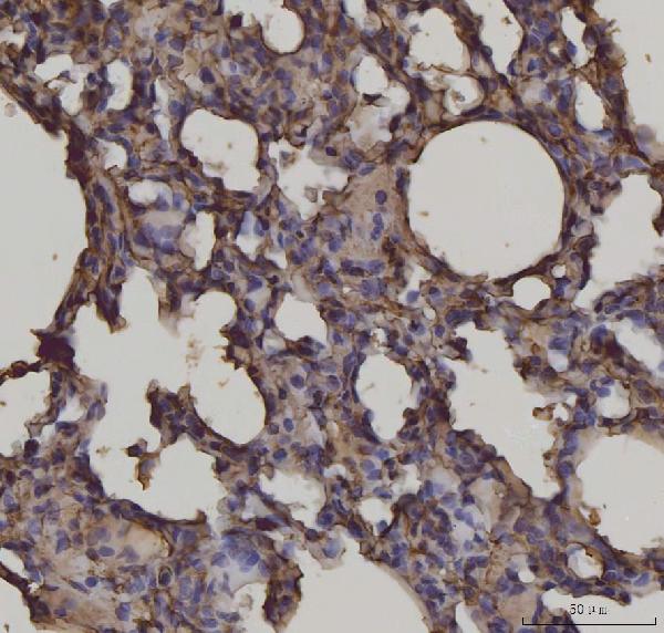
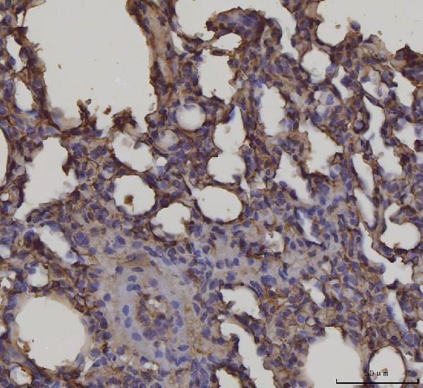
| Description | Anti-RAGE/AGER Antibody Picoband™ (monoclonal, 5C6C1) . Tested in Flow Cytometry, IHC, WB applications. This antibody reacts with Human, Mouse, Rat. |
| Reconstitution | Adding 0.2 ml of distilled water will yield a concentration of 500 µg/ml. |
| Gene ID | 177 |
|---|---|
| Other Names | Advanced glycosylation end product-specific receptor, Receptor for advanced glycosylation end products, AGER, RAGE |
| Calculated MW | 43 kDa |
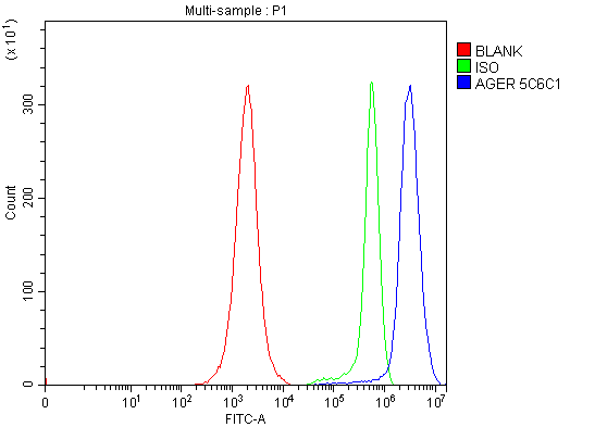
| Application Details | Western blot, 0.25-0.5 µg/ml, Mouse, Rat Immunohistochemistry(Paraffin-embedded Section), 2-5 µg/ml, Mouse, Rat Flow Cytometry, 1-3 µg/1x10^6 cells, Human |
| Contents | Each vial contains 4 mg Trehalose, 0.9 mg NaCl and 0.2 mg Na2HPO4. |
| Clone Names | Clone: 5C6C1 |
| Immunogen | A synthetic peptide corresponding to a sequence at the N-terminus of human RAGE, different from the related mouse and rat sequences by six amino acids. |
| Purification | Immunogen affinity purified. |
| Storage | At -20°C for one year from date of receipt. After reconstitution, at 4°C for one month. It can also be aliquotted and stored frozen at -20°C for six months. Avoid repeated freezing and thawing. |
| Name | AGER |
|---|---|
| Synonyms | RAGE |
| Function | Cell surface pattern recognition receptor that senses endogenous stress signals with a broad ligand repertoire including advanced glycation end products, S100 proteins, high-mobility group box 1 protein/HMGB1, amyloid beta/APP oligomers, nucleic acids, phospholipids and glycosaminoglycans (PubMed:27572515, PubMed:28515150, PubMed:34743181). Advanced glycosylation end products are nonenzymatically glycosylated proteins which accumulate in vascular tissue in aging and at an accelerated rate in diabetes (PubMed:21565706). These ligands accumulate at inflammatory sites during the pathogenesis of various diseases, including diabetes, vascular complications, neurodegenerative disorders, and cancers and RAGE transduces their binding into pro-inflammatory responses. Upon ligand binding, uses TIRAP and MYD88 as adapters to transduce the signal ultimately leading to the induction or inflammatory cytokines IL6, IL8 and TNFalpha through activation of NF-kappa-B (PubMed:21829704, PubMed:33436632). Interaction with S100A12 on endothelium, mononuclear phagocytes, and lymphocytes triggers cellular activation, with generation of key pro-inflammatory mediators (PubMed:19386136). Interaction with S100B after myocardial infarction may play a role in myocyte apoptosis by activating ERK1/2 and p53/TP53 signaling (By similarity). Contributes to the translocation of amyloid- beta peptide (ABPP) across the cell membrane from the extracellular to the intracellular space in cortical neurons (PubMed:19906677). ABPP- initiated RAGE signaling, especially stimulation of p38 mitogen- activated protein kinase (MAPK), has the capacity to drive a transport system delivering ABPP as a complex with RAGE to the intraneuronal space. Participates in endothelial albumin transcytosis together with HMGB1 through the RAGE/SRC/Caveolin-1 pathway, leading to endothelial hyperpermeability (PubMed:27572515). Mediates the loading of HMGB1 in extracellular vesicles (EVs) that shuttle HMGB1 to hepatocytes by transferrin-mediated endocytosis and subsequently promote hepatocyte pyroptosis by activating the NLRP3 inflammasome (PubMed:34743181). Promotes also extracellular hypomethylated DNA (CpG DNA) uptake by cells via the endosomal route to activate inflammatory responses (PubMed:24081950, PubMed:28515150). |
| Cellular Location | [Isoform 1]: Cell membrane; Single-pass type I membrane protein [Isoform 10]: Cell membrane; Single-pass type I membrane protein |
| Tissue Location | Endothelial cells. |

Thousands of laboratories across the world have published research that depended on the performance of antibodies from Abcepta to advance their research. Check out links to articles that cite our products in major peer-reviewed journals, organized by research category.
info@abcepta.com, and receive a free "I Love Antibodies" mug.

Provided below are standard protocols that you may find useful for product applications.
Background
The receptor for advanced glycation end products (RAGE) is a multi-ligand member of the immunoglobulin superfamily of cell surface molecules. It interacts with distinct molecules implicated in homeostasis, development and inflammation, and certain diseases such as diabetes and Alzheimer's disease. RAGE is also a central cell surface receptor for amphoterin and EN-RAGE. And RAGE is associated with sustained NF-kappaB activation in the diabetic microenvironment and has a central role in sensory neuronal dysfunction. Moreover, RAGE propagates cellular dysfunction in several inflammatory disorders and diabetes, and it also functions as an endothelial adhesion receptor promoting leukocyte recruitment.
If you have used an Abcepta product and would like to share how it has performed, please click on the "Submit Review" button and provide the requested information. Our staff will examine and post your review and contact you if needed.
If you have any additional inquiries please email technical services at tech@abcepta.com.
Ordering Information
Other Products
Shipping Information
