


 Foundational characteristics of cancer include proliferation, angiogenesis, migration, evasion of apoptosis, and cellular immortality. Find key markers for these cellular processes and antibodies to detect them.
 The SUMOplot™ Analysis Program predicts and scores sumoylation sites in your protein. SUMOylation is a post-translational modification involved in various cellular processes, such as nuclear-cytosolic transport, transcriptional regulation, apoptosis, protein stability, response to stress, and progression through the cell cycle.
 The Autophagy Receptor Motif Plotter predicts and scores autophagy receptor binding sites in your protein. Identifying proteins connected to this pathway is critical to understanding the role of autophagy in physiological as well as pathological processes such as development, differentiation, neurodegenerative diseases, stress, infection, and cancer.
Anti-XIAP Antibody Picoband™ (monoclonal, 3G2G1)
- SPECIFICATION
- CITATIONS
- PROTOCOLS
- BACKGROUND

Application
| WB |
|---|---|
| Primary Accession | P98170 |
| Host | Mouse |
| Isotype | Mouse IgG1 |
| Reactivity | Rat, Human, Mouse |
| Clonality | Monoclonal |
| Format | Lyophilized |
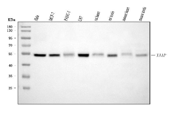
| Description | Anti-XIAP Antibody Picoband™ (monoclonal, 3G2G1) . Tested in WB applications. This antibody reacts with Human, Mouse, Rat. |
| Reconstitution | Adding 0.2 ml of distilled water will yield a concentration of 500 µg/ml. |
| Gene ID | 331 |
|---|---|
| Other Names | E3 ubiquitin-protein ligase XIAP, 2.3.2.27, Baculoviral IAP repeat-containing protein 4, IAP-like protein, ILP, hILP, Inhibitor of apoptosis protein 3, IAP-3, hIAP-3, hIAP3, RING-type E3 ubiquitin transferase XIAP, X-linked inhibitor of apoptosis protein, X-linked IAP, XIAP {ECO:0000303|PubMed:12121969, ECO:0000312|HGNC:HGNC:592} |
| Calculated MW | 54 kDa |
| Application Details | Western blot, 0.25-0.5 µg/ml, Human, Mouse, Rat |
| Contents | Each vial contains 4 mg Trehalose, 0.9 mg NaCl and 0.2 mg Na2HPO4. |
| Clone Names | Clone: 3G2G1 |
| Immunogen | E.coli-derived human XIAP recombinant protein (Position: A15-V244). Human XIAP shares 89.4% and 90.7% amino acid (aa) sequence identity with mouse and rat XIAP, respectively. |
| Purification | Immunogen affinity purified. |
| Storage | At -20°C for one year from date of receipt. After reconstitution, at 4°C for one month. It can also be aliquotted and stored frozen at -20°C for six months. Avoid repeated freezing and thawing. |
| Name | XIAP {ECO:0000303|PubMed:12121969, ECO:0000312|HGNC:HGNC:592} |
|---|---|
| Function | Multi-functional protein which regulates not only caspases and apoptosis, but also modulates inflammatory signaling and immunity, copper homeostasis, mitogenic kinase signaling, cell proliferation, as well as cell invasion and metastasis (PubMed:11257230, PubMed:11257231, PubMed:11447297, PubMed:12121969, PubMed:12620238, PubMed:17560374, PubMed:17967870, PubMed:19473982, PubMed:20154138, PubMed:22103349, PubMed:9230442). Acts as a direct caspase inhibitor (PubMed:11257230, PubMed:11257231, PubMed:12620238). Directly bind to the active site pocket of CASP3 and CASP7 and obstructs substrate entry (PubMed:11257230, PubMed:11257231, PubMed:16352606, PubMed:16916640). Inactivates CASP9 by keeping it in a monomeric, inactive state (PubMed:12620238). Acts as an E3 ubiquitin-protein ligase regulating NF-kappa-B signaling and the target proteins for its E3 ubiquitin- protein ligase activity include: RIPK1, RIPK2, MAP3K2/MEKK2, DIABLO/SMAC, AIFM1, CCS, PTEN and BIRC5/survivin (PubMed:17560374, PubMed:17967870, PubMed:19473982, PubMed:20154138, PubMed:22103349, PubMed:22607974, PubMed:29452636, PubMed:30026309). Acts as an important regulator of innate immunity by mediating 'Lys-63'-linked polyubiquitination of RIPK2 downstream of NOD1 and NOD2, thereby transforming RIPK2 into a scaffolding protein for downstream effectors, ultimately leading to activation of the NF-kappa-B and MAP kinases signaling (PubMed:19667203, PubMed:22607974, PubMed:29452636, PubMed:30026309). 'Lys-63'-linked polyubiquitination of RIPK2 also promotes recruitment of the LUBAC complex to RIPK2 (PubMed:22607974, PubMed:29452636). Regulates the BMP signaling pathway and the SMAD and MAP3K7/TAK1 dependent pathways leading to NF-kappa-B and JNK activation (PubMed:17560374). Ubiquitination of CCS leads to enhancement of its chaperone activity toward its physiologic target, SOD1, rather than proteasomal degradation (PubMed:20154138). Ubiquitination of MAP3K2/MEKK2 and AIFM1 does not lead to proteasomal degradation (PubMed:17967870, PubMed:22103349). Plays a role in copper homeostasis by ubiquitinating COMMD1 and promoting its proteasomal degradation (PubMed:14685266). Can also function as E3 ubiquitin-protein ligase of the NEDD8 conjugation pathway, targeting effector caspases for neddylation and inactivation (PubMed:21145488). Ubiquitinates and therefore mediates the proteasomal degradation of BCL2 in response to apoptosis (PubMed:29020630). Protects cells from spontaneous formation of the ripoptosome, a large multi-protein complex that has the capability to kill cancer cells in a caspase-dependent and caspase- independent manner (PubMed:22095281). Suppresses ripoptosome formation by ubiquitinating RIPK1 and CASP8 (PubMed:22095281). Acts as a positive regulator of Wnt signaling and ubiquitinates TLE1, TLE2, TLE3, TLE4 and AES (PubMed:22304967). Ubiquitination of TLE3 results in inhibition of its interaction with TCF7L2/TCF4 thereby allowing efficient recruitment and binding of the transcriptional coactivator beta-catenin to TCF7L2/TCF4 that is required to initiate a Wnt-specific transcriptional program (PubMed:22304967). |
| Cellular Location | Cytoplasm. Nucleus. Note=TLE3 promotes its nuclear localization. |
| Tissue Location | Expressed in colonic crypts (at protein level) (PubMed:30389919). Ubiquitous, except peripheral blood leukocytes (PubMed:8654366). |

Thousands of laboratories across the world have published research that depended on the performance of antibodies from Abcepta to advance their research. Check out links to articles that cite our products in major peer-reviewed journals, organized by research category.
info@abcepta.com, and receive a free "I Love Antibodies" mug.

Provided below are standard protocols that you may find useful for product applications.
Background
XIAP, also known as IAP3 or BIRC4, is a protein that stops apoptotic cell death. It is mapped to chromosome Xq25. This gene encodes a protein that belongs to a family of apoptotic suppressor proteins. Members of this family share a conserved motif termed, baculovirus IAP repeat, which is necessary for their anti-apoptotic function. This protein functions through binding to tumor necrosis factor receptor-associated factors TRAF1 and TRAF2 and inhibits apoptosis induced by menadione, a potent inducer of free radicals, and interleukin 1-beta converting enzyme. This protein also inhibits at least two members of the caspase family of cell-death proteases, caspase-3 and caspase-7. Mutations in this gene are the cause of X-linked lymphoproliferative syndrome. Alternate splicing results in multiple transcript variants.
If you have used an Abcepta product and would like to share how it has performed, please click on the "Submit Review" button and provide the requested information. Our staff will examine and post your review and contact you if needed.
If you have any additional inquiries please email technical services at tech@abcepta.com.
Ordering Information
Other Products
Shipping Information
