


 Foundational characteristics of cancer include proliferation, angiogenesis, migration, evasion of apoptosis, and cellular immortality. Find key markers for these cellular processes and antibodies to detect them.
 The SUMOplot™ Analysis Program predicts and scores sumoylation sites in your protein. SUMOylation is a post-translational modification involved in various cellular processes, such as nuclear-cytosolic transport, transcriptional regulation, apoptosis, protein stability, response to stress, and progression through the cell cycle.
 The Autophagy Receptor Motif Plotter predicts and scores autophagy receptor binding sites in your protein. Identifying proteins connected to this pathway is critical to understanding the role of autophagy in physiological as well as pathological processes such as development, differentiation, neurodegenerative diseases, stress, infection, and cancer.
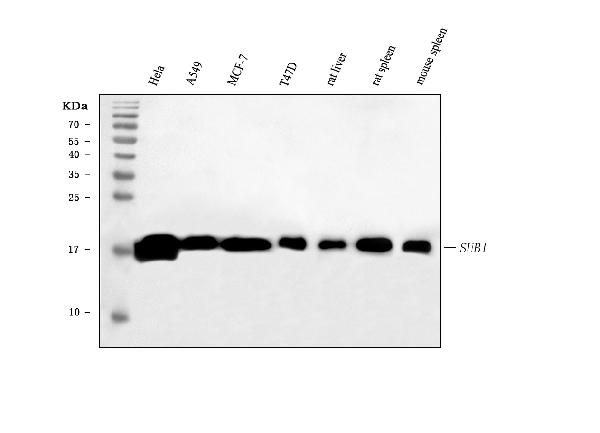
Anti-PC4/SUB1 Antibody Picoband™ (monoclonal, 8D9D1)
- SPECIFICATION
- CITATIONS
- PROTOCOLS
- BACKGROUND

Application
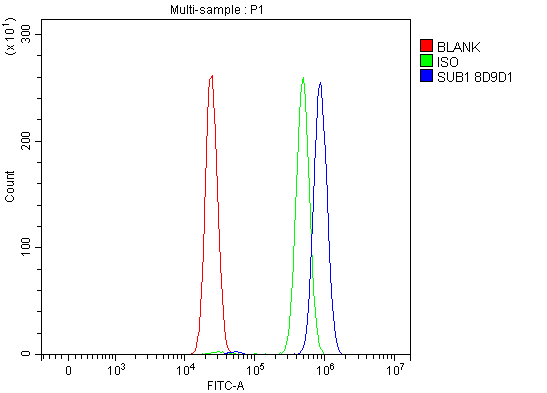
| WB, IHC, IF, FC |
|---|---|
| Primary Accession | P53999 |
| Host | Mouse |
| Isotype | Mouse IgG2b |
| Reactivity | Rat, Human, Mouse |
| Clonality | Monoclonal |
| Format | Lyophilized |
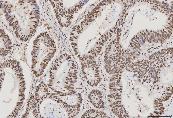
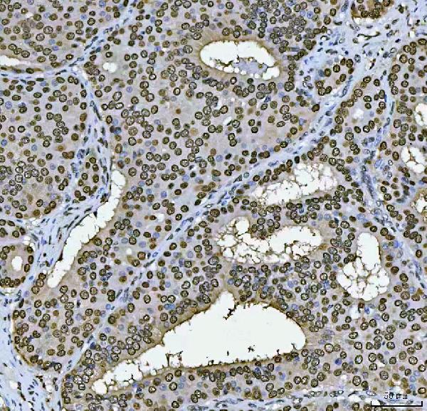
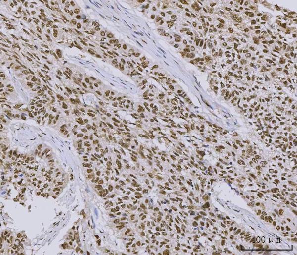
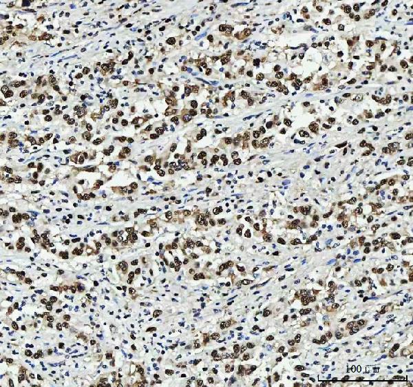
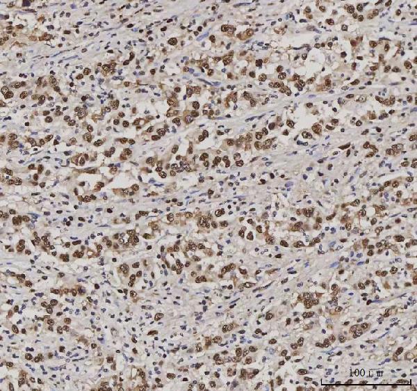
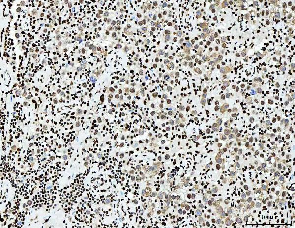
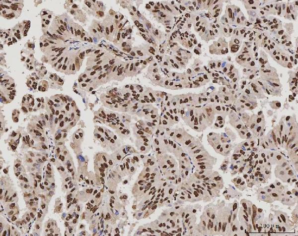
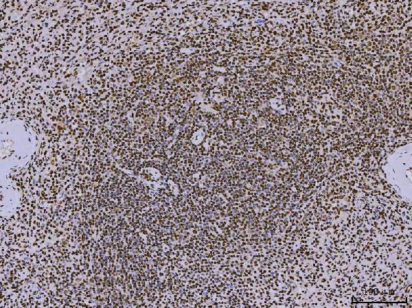
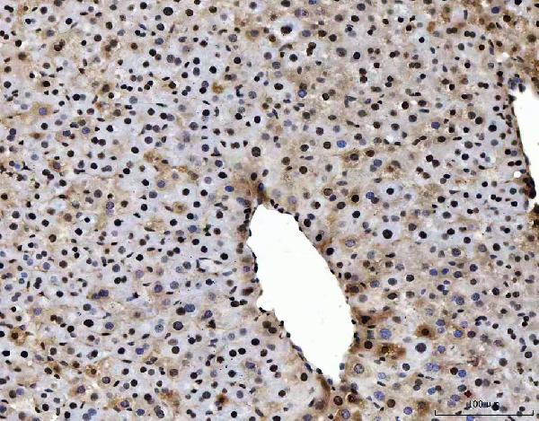
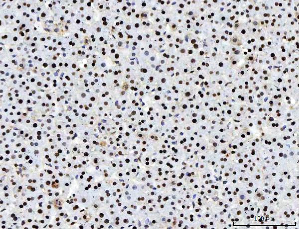
| Description | Anti-PC4/SUB1 Antibody Picoband™ (monoclonal, 8D9D1) . Tested in Flow Cytometry, IF, IHC, WB applications. This antibody reacts with Human, Mouse, Rat. |
| Reconstitution | Adding 0.2 ml of distilled water will yield a concentration of 500 µg/ml. |
| Gene ID | 10923 |
|---|---|
| Other Names | Activated RNA polymerase II transcriptional coactivator p15, Positive cofactor 4, PC4, SUB1 homolog, p14, SUB1, PC4, RPO2TC1 |
| Calculated MW | 19 kDa |
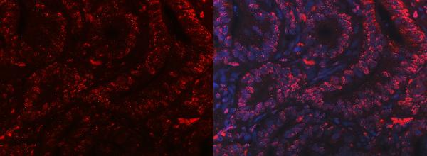
| Application Details | Western blot, 0.25-0.5 µg/ml, Human, Mouse, Rat Immunohistochemistry(Paraffin-embedded Section), 2-5 µg/ml, Human, Mouse, Rat Immunofluorescence, 5 µg/ml, Human Flow Cytometry, 1-3 µg/1x10^6 cells, Human |
| Contents | Each vial contains 4 mg Trehalose, 0.9 mg NaCl and 0.2 mg Na2HPO4. |
| Clone Names | Clone: 8D9D1 |
| Immunogen | E.coli-derived human PC4/SUB1 recombinant protein (Position: N62-L127). |
| Purification | Immunogen affinity purified. |
| Storage | At -20°C for one year from date of receipt. After reconstitution, at 4°C for one month. It can also be aliquotted and stored frozen at -20°C for six months. Avoid repeated freezing and thawing. |
| Name | SUB1 |
|---|---|
| Synonyms | PC4, RPO2TC1 |
| Function | General coactivator that functions cooperatively with TAFs and mediates functional interactions between upstream activators and the general transcriptional machinery. May be involved in stabilizing the multiprotein transcription complex. Binds single-stranded DNA. Also binds, in vitro, non-specifically to double-stranded DNA (ds DNA). |
| Cellular Location | Nucleus. |

Thousands of laboratories across the world have published research that depended on the performance of antibodies from Abcepta to advance their research. Check out links to articles that cite our products in major peer-reviewed journals, organized by research category.
info@abcepta.com, and receive a free "I Love Antibodies" mug.

Provided below are standard protocols that you may find useful for product applications.
Background
Activated RNA polymerase II transcriptional coactivator p15, also known as positive cofactor 4 (PC4) or SUB1 homolog, is a protein that in humans is encoded by the SUB1 gene. This gene is mapped to 5p13.3. The transcriptional cofactor PC4 is an ancient single-strand DNA (ssDNA)-binding protein that has a homologue in bacteriophage T5 where it is likely the elusive replicative ssDNA-binding protein. The recombinant PC4 is shown to function identically to the native protein through its interaction with TAFs.
If you have used an Abcepta product and would like to share how it has performed, please click on the "Submit Review" button and provide the requested information. Our staff will examine and post your review and contact you if needed.
If you have any additional inquiries please email technical services at tech@abcepta.com.
Ordering Information
Other Products
Shipping Information
