


 Foundational characteristics of cancer include proliferation, angiogenesis, migration, evasion of apoptosis, and cellular immortality. Find key markers for these cellular processes and antibodies to detect them.
 The SUMOplot™ Analysis Program predicts and scores sumoylation sites in your protein. SUMOylation is a post-translational modification involved in various cellular processes, such as nuclear-cytosolic transport, transcriptional regulation, apoptosis, protein stability, response to stress, and progression through the cell cycle.
 The Autophagy Receptor Motif Plotter predicts and scores autophagy receptor binding sites in your protein. Identifying proteins connected to this pathway is critical to understanding the role of autophagy in physiological as well as pathological processes such as development, differentiation, neurodegenerative diseases, stress, infection, and cancer.
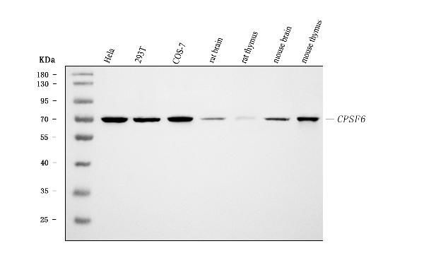
Anti-CPSF6 Antibody Picoband™ (monoclonal, 3F11E1)


- SPECIFICATION
- CITATIONS
- PROTOCOLS
- BACKGROUND

Application
| WB, IHC, IF, ICC, FC |
|---|---|
| Primary Accession | Q16630 |
| Host | Mouse |
| Isotype | Mouse IgG1 |
| Reactivity | Rat, Human, Mouse, Monkey |
| Clonality | Monoclonal |
| Format | Lyophilized |
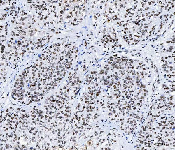
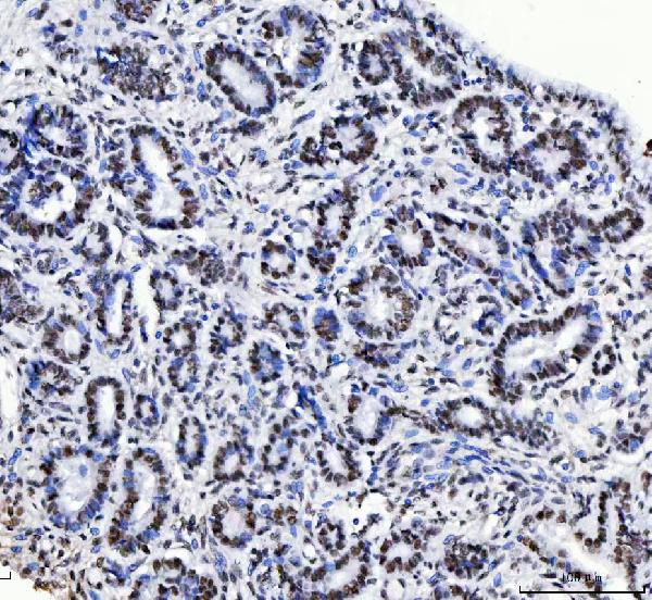
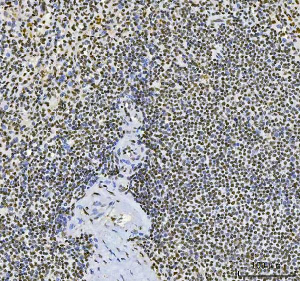
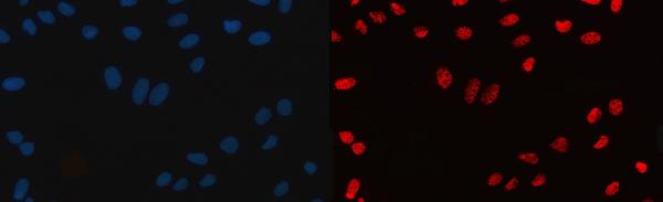
| Description | Anti-CPSF6 Antibody Picoband™ (monoclonal, 3F11E1) . Tested in Flow Cytometry, IF, IHC, ICC, WB applications. This antibody reacts with Human, Mouse, Rat, Monkey. |
| Reconstitution | Adding 0.2 ml of distilled water will yield a concentration of 500 µg/ml. |
| Gene ID | 11052 |
|---|---|
| Other Names | Cleavage and polyadenylation specificity factor subunit 6, Cleavage and polyadenylation specificity factor 68 kDa subunit, CPSF 68 kDa subunit, Cleavage factor Im complex 68 kDa subunit, CFIm68, Pre-mRNA cleavage factor Im 68 kDa subunit, Protein HPBRII-4/7, CPSF6 (HGNC:13871) |
| Calculated MW | 68 kDa |
| Application Details | Western blot, 0.25-0.5 µg/ml, Human, Mouse, Rat, Monkey Immunohistochemistry(Paraffin-embedded Section), 2-5 µg/ml, Human, Mouse, Rat Immunocytochemistry/Immunofluorescence, 5 µg/ml, Human Flow Cytometry, 1-3 µg/1x10^6 cells, Human |
| Contents | Each vial contains 4 mg Trehalose, 0.9 mg NaCl and 0.2 mg Na2HPO4. |
| Clone Names | Clone: 3F11E1 |
| Immunogen | E.coli-derived human CPSF6 recombinant protein (Position: R50-Q176). |
| Purification | Immunogen affinity purified. |
| Storage | At -20°C for one year from date of receipt. After reconstitution, at 4°C for one month. It can also be aliquotted and stored frozen at -20°C for six months. Avoid repeated freezing and thawing. |
| Name | CPSF6 (HGNC:13871) |
|---|---|
| Function | Component of the cleavage factor Im (CFIm) complex that functions as an activator of the pre-mRNA 3'-end cleavage and polyadenylation processing required for the maturation of pre-mRNA into functional mRNAs (PubMed:14690600, PubMed:29276085, PubMed:8626397, PubMed:9659921). CFIm contributes to the recruitment of multiprotein complexes on specific sequences on the pre-mRNA 3'-end, so called cleavage and polyadenylation signals (pA signals) (PubMed:14690600, PubMed:8626397, PubMed:9659921). Most pre-mRNAs contain multiple pA signals, resulting in alternative cleavage and polyadenylation (APA) producing mRNAs with variable 3'-end formation (PubMed:23187700, PubMed:29276085). The CFIm complex acts as a key regulator of cleavage and polyadenylation site choice during APA through its binding to 5'- UGUA-3' elements localized in the 3'-untranslated region (UTR) for a huge number of pre-mRNAs (PubMed:20695905, PubMed:29276085). CPSF6 enhances NUDT21/CPSF5 binding to 5'-UGUA-3' elements localized upstream of pA signals and promotes RNA looping, and hence activates directly the mRNA 3'-processing machinery (PubMed:15169763, PubMed:21295486, PubMed:29276085). Plays a role in mRNA export (PubMed:19864460). |
| Cellular Location | Nucleus. Nucleus, nucleoplasm. Nucleus speckle. Cytoplasm. Note=Shuttles between the nucleus and the cytoplasm in a transcription- and XPO1/CRM1-independent manner, most probably in complex with the cleavage factor Im complex (CFIm) (PubMed:19864460). Colocalizes with PSPC1 in punctate subnuclear structures often located adjacent to nuclear speckles, called paraspeckles, and corresponding to interchromatin granules-associated zones (IGAZs) (PubMed:17267687). Distribution in speckles and paraspeckles varies during the cell cycle (PubMed:17267687). Associates at sites of active transcription on nascent perichromatin fibrils (PFs) and perichromatin granules (PubMed:17267687). Nuclear import is mediated via interaction with TNPO3 independently of CPSF6 phosphorylation status (PubMed:30916345). |

Thousands of laboratories across the world have published research that depended on the performance of antibodies from Abcepta to advance their research. Check out links to articles that cite our products in major peer-reviewed journals, organized by research category.
info@abcepta.com, and receive a free "I Love Antibodies" mug.

Provided below are standard protocols that you may find useful for product applications.
Background
Cleavage and polyadenylation specificity factor subunit 6 is a protein that in humans is encoded by the CPSF6 gene. The protein encoded by this gene is one subunit of a cleavage factor required for 3' RNA cleavage and polyadenylation processing. The interaction of the protein with the RNA is one of the earliest steps in the assembly of the 3' end processing complex and facilitates the recruitment of other processing factors. The cleavage factor complex is composed of four polypeptides. This gene encodes the 68kD subunit. It has a domain organization reminiscent of spliceosomal proteins.
If you have used an Abcepta product and would like to share how it has performed, please click on the "Submit Review" button and provide the requested information. Our staff will examine and post your review and contact you if needed.
If you have any additional inquiries please email technical services at tech@abcepta.com.
Ordering Information
Other Products
Shipping Information
