


 Foundational characteristics of cancer include proliferation, angiogenesis, migration, evasion of apoptosis, and cellular immortality. Find key markers for these cellular processes and antibodies to detect them.
 The SUMOplot™ Analysis Program predicts and scores sumoylation sites in your protein. SUMOylation is a post-translational modification involved in various cellular processes, such as nuclear-cytosolic transport, transcriptional regulation, apoptosis, protein stability, response to stress, and progression through the cell cycle.
 The Autophagy Receptor Motif Plotter predicts and scores autophagy receptor binding sites in your protein. Identifying proteins connected to this pathway is critical to understanding the role of autophagy in physiological as well as pathological processes such as development, differentiation, neurodegenerative diseases, stress, infection, and cancer.
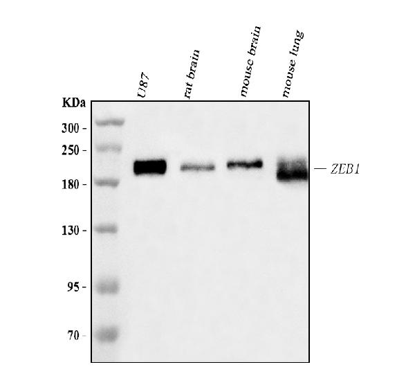
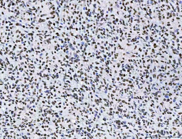
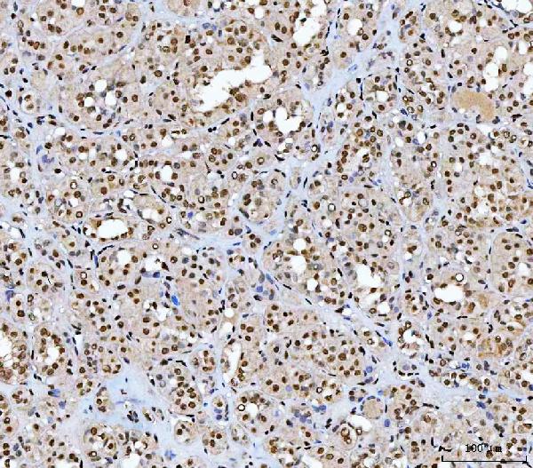
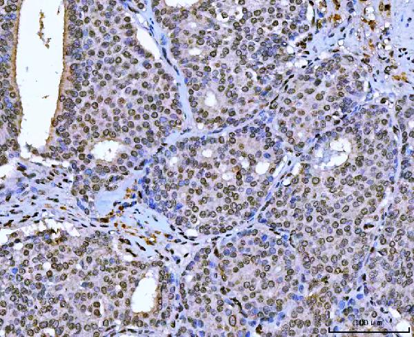
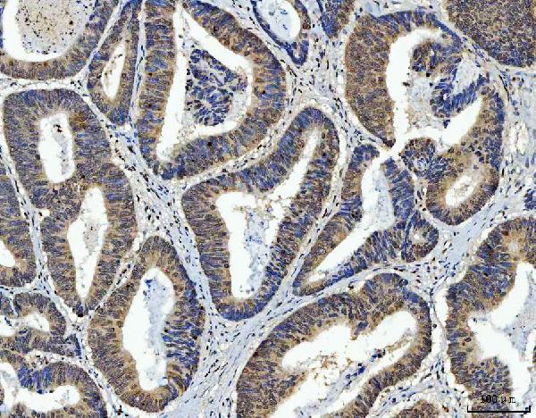
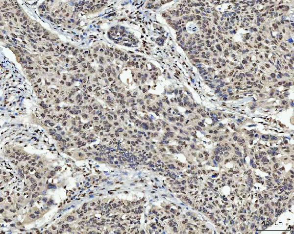
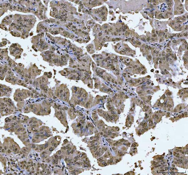
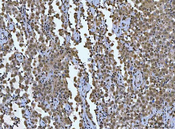
Anti-AREB6/ZEB1 Antibody Picoband™ (monoclonal, 8B12D7)
- SPECIFICATION
- CITATIONS
- PROTOCOLS
- BACKGROUND

Application
| WB, IHC, IF, ICC |
|---|---|
| Primary Accession | P37275 |
| Host | Mouse |
| Isotype | Mouse IgG2b |
| Reactivity | Rat, Human, Mouse |
| Clonality | Monoclonal |
| Format | Lyophilized |
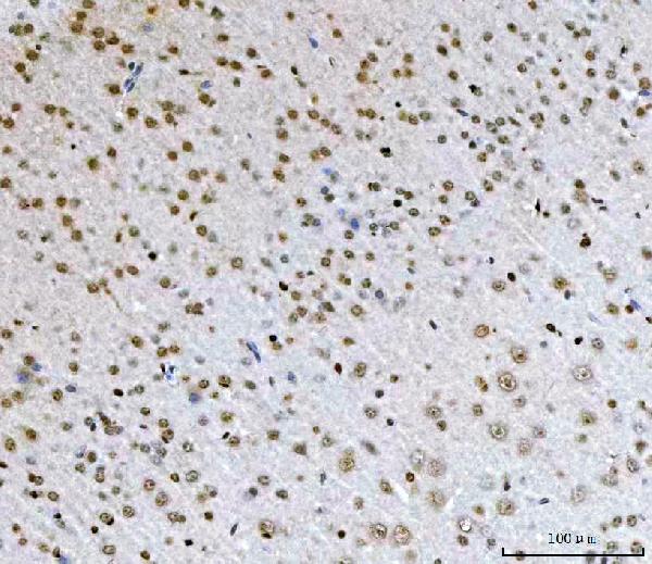
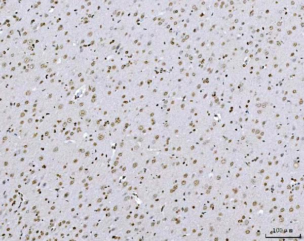
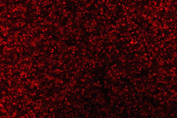
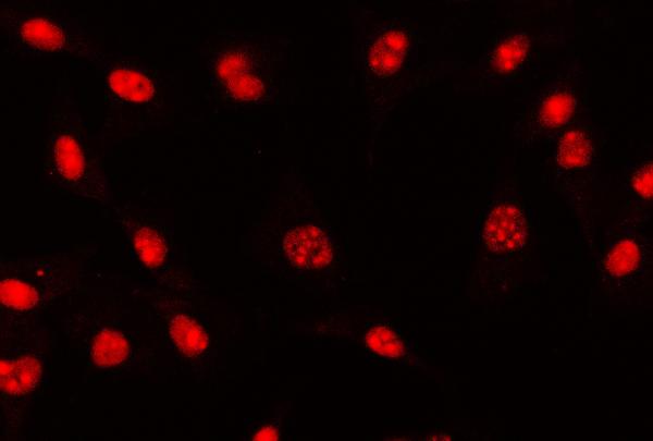
| Description | Anti-AREB6/ZEB1 Antibody Picoband™ (monoclonal, 8B12D7) . Tested in IF, IHC, ICC, WB applications. This antibody reacts with Human, Mouse, Rat. |
| Reconstitution | Adding 0.2 ml of distilled water will yield a concentration of 500 µg/ml. |
| Gene ID | 6935 |
|---|---|
| Other Names | Zinc finger E-box-binding homeobox 1 {ECO:0000312|HGNC:HGNC:11642}, NIL-2-A zinc finger protein {ECO:0000303|Ref.26}, Negative regulator of IL2, Transcription factor 8, TCF-8, ZEB1 (HGNC:11642) |
| Calculated MW | 200 kDa |
| Application Details | Western blot, 0.25-0.5 µg/ml, Human, Mouse, Rat Immunohistochemistry(Paraffin-embedded Section), 2-5 µg/ml, Human, Mouse, Rat Immunocytochemistry/Immunofluorescence, 5 µg/ml, Human Immunofluorescence, 5 µg/ml, Human |
| Contents | Each vial contains 4 mg Trehalose, 0.9 mg NaCl and 0.2 mg Na2HPO4. |
| Clone Names | Clone: 8B12D7 |
| Immunogen | A synthetic peptide corresponding to a sequence in the middle region of human AREB6/ZEB1. |
| Purification | Immunogen affinity purified. |
| Storage | At -20°C for one year from date of receipt. After reconstitution, at 4°C for one month. It can also be aliquotted and stored frozen at -20°C for six months. Avoid repeated freezing and thawing. |
| Name | ZEB1 (HGNC:11642) |
|---|---|
| Function | Acts as a transcriptional repressor. Inhibits interleukin-2 (IL-2) gene expression. Enhances or represses the promoter activity of the ATP1A1 gene depending on the quantity of cDNA and on the cell type. Represses E-cadherin promoter and induces an epithelial-mesenchymal transition (EMT) by recruiting SMARCA4/BRG1. Represses BCL6 transcription in the presence of the corepressor CTBP1. Positively regulates neuronal differentiation. Represses RCOR1 transcription activation during neurogenesis. Represses transcription by binding to the E box (5'-CANNTG-3'). In the absence of TGFB1, acts as a repressor of COL1A2 transcription via binding to the E-box in the upstream enhancer region (By similarity). |
| Cellular Location | Nucleus |
| Tissue Location | Colocalizes with SMARCA4/BRG1 in E-cadherin- negative cells from established lines, and stroma of normal colon as well as in de-differentiated epithelial cells at the invasion front of colorectal carcinomas (at protein level). Expressed in heart and skeletal muscle, but not in liver, spleen, or pancreas |

Thousands of laboratories across the world have published research that depended on the performance of antibodies from Abcepta to advance their research. Check out links to articles that cite our products in major peer-reviewed journals, organized by research category.
info@abcepta.com, and receive a free "I Love Antibodies" mug.

Provided below are standard protocols that you may find useful for product applications.
Background
ZEB1 (Zinc Finger E Box-Binding Homeobox 1), also called TCF8, NIL2A or DELTA-EF1, is a protein that in humans is encoded by the ZEB1 gene. Fluorescence in situ hybridization localized the ZEB1 gene to chromosome 10p11.2. Krafchak et al. (2005) demonstrated a complex (core plus secondary) binding site for TCF8 in the promoter of the COL4A3 gene, mutant in Alport syndrome and which encodes collagen type IV alpha-3. They detected expression of TCF8 in cornea. Nishimura et al. (2006) found that delta-Ef1 was upregulated during differentiation in a mouse smooth muscle cell (SMC) line.
If you have used an Abcepta product and would like to share how it has performed, please click on the "Submit Review" button and provide the requested information. Our staff will examine and post your review and contact you if needed.
If you have any additional inquiries please email technical services at tech@abcepta.com.
Ordering Information
Other Products
Shipping Information
