


 Foundational characteristics of cancer include proliferation, angiogenesis, migration, evasion of apoptosis, and cellular immortality. Find key markers for these cellular processes and antibodies to detect them.
 The SUMOplot™ Analysis Program predicts and scores sumoylation sites in your protein. SUMOylation is a post-translational modification involved in various cellular processes, such as nuclear-cytosolic transport, transcriptional regulation, apoptosis, protein stability, response to stress, and progression through the cell cycle.
 The Autophagy Receptor Motif Plotter predicts and scores autophagy receptor binding sites in your protein. Identifying proteins connected to this pathway is critical to understanding the role of autophagy in physiological as well as pathological processes such as development, differentiation, neurodegenerative diseases, stress, infection, and cancer.
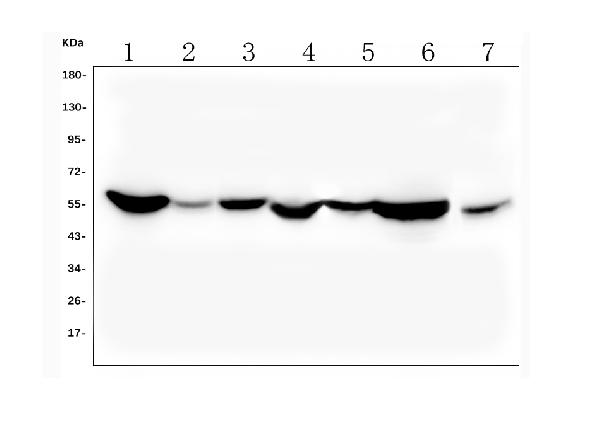
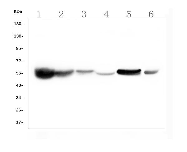
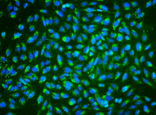
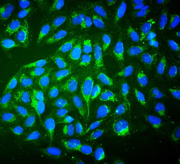
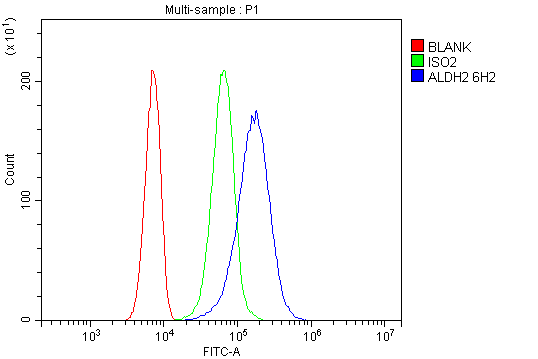
Anti-ALDH2 Antibody Picoband™(monoclonal, 6H2)
- SPECIFICATION
- CITATIONS
- PROTOCOLS
- BACKGROUND

Application
| WB, IF, ICC, FC |
|---|---|
| Primary Accession | P05091 |
| Host | Mouse |
| Isotype | Mouse IgG1 |
| Reactivity | Rat, Human, Mouse |
| Clonality | Monoclonal |
| Format | Lyophilized |
| Description | Anti-ALDH2 Antibody Picoband™ (monoclonal, 6H2) . Tested in Flow Cytometry, IF, ICC, WB applications. This antibody reacts with Human, Mouse, Rat. |
| Reconstitution | Add 0.2ml of distilled water will yield a concentration of 500 µg/ml. |
| Gene ID | 217 |
|---|---|
| Other Names | Aldehyde dehydrogenase, mitochondrial, 1.2.1.3, ALDH class 2, ALDH-E2, ALDHI, ALDH2, ALDM |
| Calculated MW | 56381 Da |
| Application Details | Western blot, 0.1-0.5 µg/ml, Human, Mouse, Rat Immunocytochemistry/Immunofluorescence, 2 µg/ml, Human Flow Cytometry, 1-3 µg/1x10^6 cells, Human |
| Contents | Each vial contains 4 mg Trehalose, 0.9 mg NaCl, 0.2 mg Na2HPO4, 0.05 mg NaN3. |
| Clone Names | Clone: 6H2 |
| Immunogen | A synthetic peptide corresponding to a sequence at the N-terminus of human ALDH2, different from the related mouse sequence by two amino acids, and from the related rat sequence by one amino acid. |
| Purification | Immunogen affinity purified. |
| Storage | Store at -20˚C for one year from date of receipt. After reconstitution, at 4˚C for one month. It can also be aliquotted and stored frozen at -20˚C for six months. Avoid repeated freeze-thaw cycles. |
| Name | ALDH2 |
|---|---|
| Synonyms | ALDM |
| Function | Required for clearance of cellular formaldehyde, a cytotoxic and carcinogenic metabolite that induces DNA damage. |
| Cellular Location | Mitochondrion matrix. |

Thousands of laboratories across the world have published research that depended on the performance of antibodies from Abcepta to advance their research. Check out links to articles that cite our products in major peer-reviewed journals, organized by research category.
info@abcepta.com, and receive a free "I Love Antibodies" mug.

Provided below are standard protocols that you may find useful for product applications.
Background
ALDH2 (Aldehyde Dehydrogenase 2 Family) is a human gene. The enzyme encoded by this gene belongs to the aldehyde dehydrogenase family of enzymes that catalyze the chemical transformation from acetaldehyde to acetic acid. Aldehyde dehydrogenase is the second enzyme of the major oxidative pathway of alcohol metabolism. Hsu et al. (1985) assigned the ALDH2 locus to chromosome 12 by means of a cDNA probe and Southern blot analysis of somatic cell hybrids. Using an unbiased proteomic search, Chen et al. (2008) identified mitochondrial ALDH2 as an enzyme whose activation correlated with reduced ischemic heart damage in rodent models. A high-throughput screen identified a small molecule activator of ALDH2, which they called Alda-1, that, when administered to rats before an ischemic event, reduced infarct size by 60%, most likely through its inhibitory effect on the formation of cytotoxic aldehydes.
If you have used an Abcepta product and would like to share how it has performed, please click on the "Submit Review" button and provide the requested information. Our staff will examine and post your review and contact you if needed.
If you have any additional inquiries please email technical services at tech@abcepta.com.
Ordering Information
Other Products
Shipping Information
