


 Foundational characteristics of cancer include proliferation, angiogenesis, migration, evasion of apoptosis, and cellular immortality. Find key markers for these cellular processes and antibodies to detect them.
 The SUMOplot™ Analysis Program predicts and scores sumoylation sites in your protein. SUMOylation is a post-translational modification involved in various cellular processes, such as nuclear-cytosolic transport, transcriptional regulation, apoptosis, protein stability, response to stress, and progression through the cell cycle.
 The Autophagy Receptor Motif Plotter predicts and scores autophagy receptor binding sites in your protein. Identifying proteins connected to this pathway is critical to understanding the role of autophagy in physiological as well as pathological processes such as development, differentiation, neurodegenerative diseases, stress, infection, and cancer.
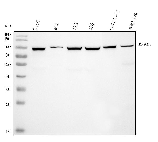
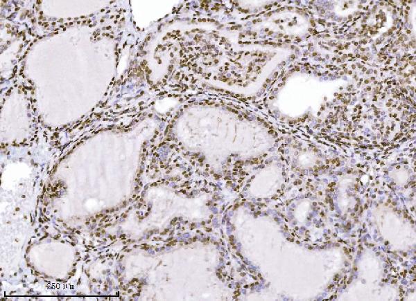
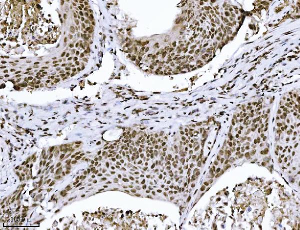
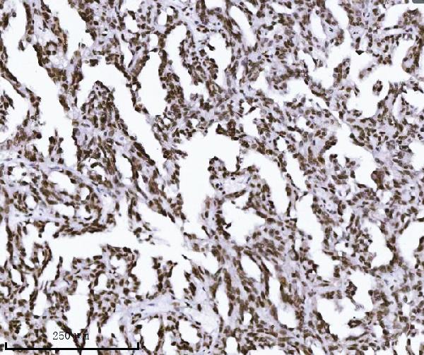
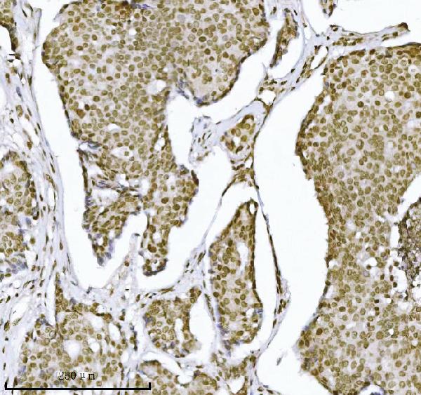
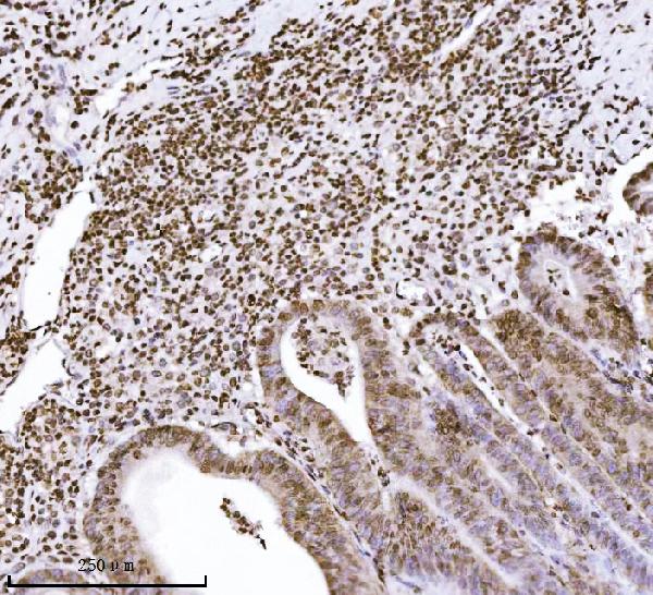
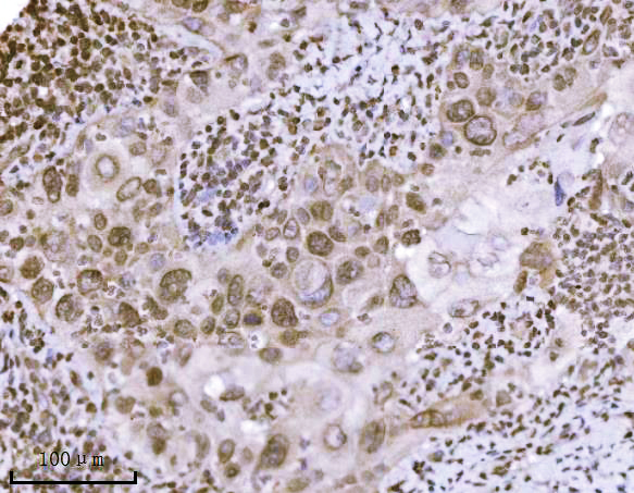
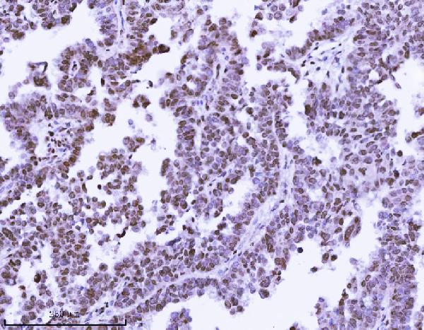
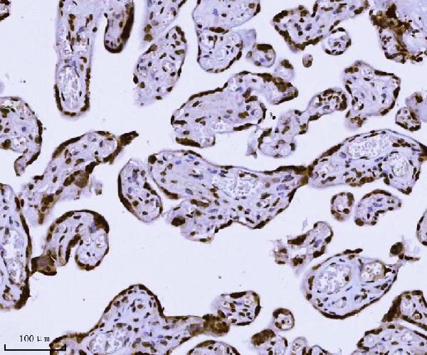
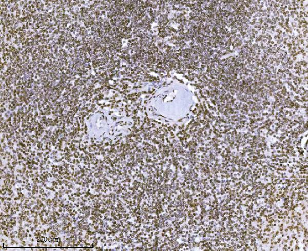
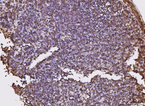
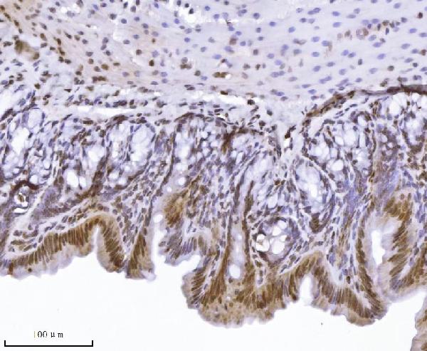
Anti-KPNB1 Antibody Picoband™ (monoclonal, 3I11F2)
- SPECIFICATION
- CITATIONS
- PROTOCOLS
- BACKGROUND

Application
| WB, IHC, IF, ICC, FC |
|---|---|
| Primary Accession | Q14974 |
| Host | Mouse |
| Isotype | Mouse IgG2b |
| Reactivity | Rat, Human, Mouse |
| Clonality | Monoclonal |
| Format | Lyophilized |
| Description | Anti-KPNB1 Antibody Picoband™ (monoclonal, 3I11F2) . Tested in Flow Cytometry, IF, IHC, ICC, WB applications. This antibody reacts with Human, Mouse, Rat. |
| Reconstitution | Adding 0.2 ml of distilled water will yield a concentration of 500 µg/ml. |
| Gene ID | 3837 |
|---|---|
| Other Names | Importin subunit beta-1, Importin-90, Karyopherin subunit beta-1, Nuclear factor p97, Pore targeting complex 97 kDa subunit, PTAC97, KPNB1, NTF97 |
| Calculated MW | 97 kDa |
| Application Details | Western blot, 0.25-0.5 µg/ml, Human, Mouse Immunohistochemistry(Paraffin-embedded Section), 2-5 µg/ml, Human, Mouse, Rat Immunocytochemistry/Immunofluorescence, 5 µg/ml, Human Flow Cytometry, 1-3 µg/1x10^6 cells, Human |
| Contents | Each vial contains 4 mg Trehalose, 0.9 mg NaCl and 0.2 mg Na2HPO4. |
| Clone Names | Clone: 3I11F2 |
| Immunogen | E.coli-derived human KPNB1 recombinant protein (Position: E8-A876). |
| Purification | Immunogen affinity purified. |
| Storage | At -20°C for one year from date of receipt. After reconstitution, at 4°C for one month. It can also be aliquotted and stored frozen at -20°C for six months. Avoid repeated freezing and thawing. |
| Name | KPNB1 |
|---|---|
| Synonyms | NTF97 |
| Function | Functions in nuclear protein import, either in association with an adapter protein, like an importin-alpha subunit, which binds to nuclear localization signals (NLS) in cargo substrates, or by acting as autonomous nuclear transport receptor (PubMed:10228156, PubMed:11682607, PubMed:11891849, PubMed:19386897, PubMed:20818336, PubMed:24699649, PubMed:7615630, PubMed:9687515). Acting autonomously, serves itself as NLS receptor (PubMed:10228156, PubMed:11682607, PubMed:11891849, PubMed:19386897, PubMed:20818336, PubMed:24699649, PubMed:7615630, PubMed:9687515). Docking of the importin/substrate complex to the nuclear pore complex (NPC) is mediated by KPNB1 through binding to nucleoporin FxFG repeats and the complex is subsequently translocated through the pore by an energy requiring, Ran-dependent mechanism (PubMed:10228156, PubMed:11682607, PubMed:11891849, PubMed:19386897, PubMed:20818336, PubMed:24699649, PubMed:7615630, PubMed:9687515). At the nucleoplasmic side of the NPC, Ran binds to importin-beta and the three components separate and importin-alpha and -beta are re-exported from the nucleus to the cytoplasm where GTP hydrolysis releases Ran from importin (PubMed:10228156, PubMed:11682607, PubMed:11891849, PubMed:19386897, PubMed:20818336, PubMed:24699649, PubMed:7615630, PubMed:9687515). The directionality of nuclear import is thought to be conferred by an asymmetric distribution of the GTP- and GDP-bound forms of Ran between the cytoplasm and nucleus (PubMed:10228156, PubMed:11682607, PubMed:11891849, PubMed:19386897, PubMed:24699649, PubMed:7615630, PubMed:9687515). Mediates autonomously the nuclear import of ribosomal proteins RPL23A, RPS7 and RPL5 (PubMed:11682607, PubMed:9687515). In association with IPO7, mediates the nuclear import of H1 histone (PubMed:10228156). In vitro, mediates nuclear import of H2A, H2B, H3 and H4 histones (By similarity). Imports MRTFA, SNAI1 and PRKCI into the nucleus (PubMed:11891849, PubMed:19386897, PubMed:20818336, PubMed:24699649). |
| Cellular Location | Cytoplasm. Nucleus envelope |

Thousands of laboratories across the world have published research that depended on the performance of antibodies from Abcepta to advance their research. Check out links to articles that cite our products in major peer-reviewed journals, organized by research category.
info@abcepta.com, and receive a free "I Love Antibodies" mug.

Provided below are standard protocols that you may find useful for product applications.
Background
Importin subunit beta-1 is a protein that in humans is encoded by the KPNB1 gene. Nucleocytoplasmic transport, a signal- and energy-dependent process, takes place through nuclear pore complexes embedded in the nuclear envelope. The import of proteins containing a nuclear localization signal (NLS) requires the NLS import receptor, a heterodimer of importin alpha and beta subunits also known as karyopherins. Importin alpha binds the NLS-containing cargo in the cytoplasm and importin beta docks the complex at the cytoplasmic side of the nuclear pore complex. In the presence of nucleoside triphosphates and the small GTP binding protein Ran, the complex moves into the nuclear pore complex and the importin subunits dissociate. Importin alpha enters the nucleoplasm with its passenger protein and importin beta remains at the pore. Interactions between importin beta and the FG repeats of nucleoporins are essential in translocation through the pore complex. The protein encoded by this gene is a member of the importin beta family. Two transcript variants encoding different isoforms have been found for this gene.
If you have used an Abcepta product and would like to share how it has performed, please click on the "Submit Review" button and provide the requested information. Our staff will examine and post your review and contact you if needed.
If you have any additional inquiries please email technical services at tech@abcepta.com.
Ordering Information
Other Products
Shipping Information
