


 Foundational characteristics of cancer include proliferation, angiogenesis, migration, evasion of apoptosis, and cellular immortality. Find key markers for these cellular processes and antibodies to detect them.
 The SUMOplot™ Analysis Program predicts and scores sumoylation sites in your protein. SUMOylation is a post-translational modification involved in various cellular processes, such as nuclear-cytosolic transport, transcriptional regulation, apoptosis, protein stability, response to stress, and progression through the cell cycle.
 The Autophagy Receptor Motif Plotter predicts and scores autophagy receptor binding sites in your protein. Identifying proteins connected to this pathway is critical to understanding the role of autophagy in physiological as well as pathological processes such as development, differentiation, neurodegenerative diseases, stress, infection, and cancer.
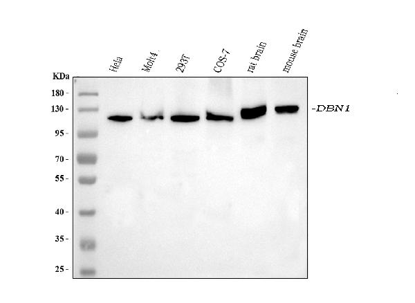
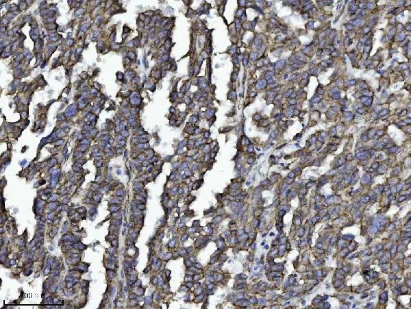
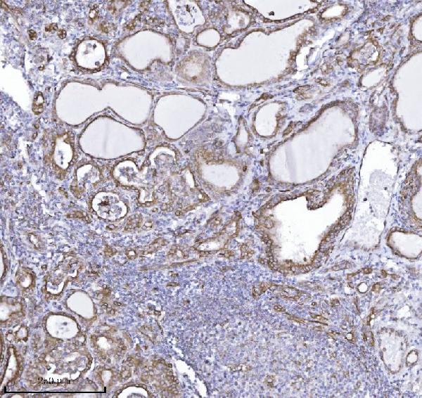
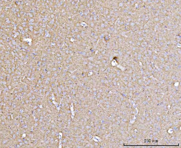
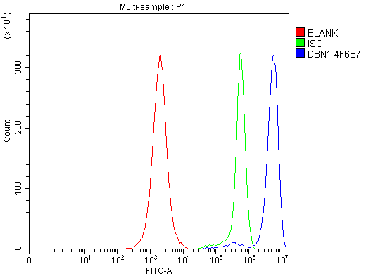
Anti-Drebrin/DBN1 Antibody Picoband™ (monoclonal, 4F6E7)
- SPECIFICATION
- CITATIONS
- PROTOCOLS
- BACKGROUND

Application
| WB, IHC, FC |
|---|---|
| Primary Accession | Q16643 |
| Host | Mouse |
| Isotype | Mouse IgG2a |
| Reactivity | Rat, Human, Mouse, Monkey |
| Clonality | Monoclonal |
| Format | Lyophilized |
| Description | Anti-Drebrin/DBN1 Antibody Picoband™ (monoclonal, 4F6E7) . Tested in Flow Cytometry, IHC, WB applications. This antibody reacts with Human, Mouse, Rat, Monkey. |
| Reconstitution | Adding 0.2 ml of distilled water will yield a concentration of 500 µg/ml. |
| Gene ID | 1627 |
|---|---|
| Other Names | Drebrin, Developmentally-regulated brain protein, DBN1, D0S117E |
| Calculated MW | 120 kDa |
| Application Details | Western blot, 0.25-0.5 µg/ml, Human, Mouse, Rat, Monkey Immunohistochemistry(Paraffin-embedded Section), 2-5 µg/ml, Human, Rat Flow Cytometry, 1-3 µg/1x10^6 cells, Human |
| Contents | Each vial contains 4 mg Trehalose, 0.9 mg NaCl and 0.2 mg Na2HPO4. |
| Clone Names | Clone: 4F6E7 |
| Immunogen | E.coli-derived human Drebrin/DBN1 recombinant protein (Position: H9-D649). |
| Purification | Immunogen affinity purified. |
| Storage | At -20°C for one year from date of receipt. After reconstitution, at 4°C for one month. It can also be aliquotted and stored frozen at -20°C for six months. Avoid repeated freezing and thawing. |
| Name | DBN1 |
|---|---|
| Synonyms | D0S117E |
| Function | Actin cytoskeleton-organizing protein that plays a role in the formation of cell projections (PubMed:20215400). Required for actin polymerization at immunological synapses (IS) and for the recruitment of the chemokine receptor CXCR4 to IS (PubMed:20215400). Plays a role in dendritic spine morphogenesis and organization, including the localization of the dopamine receptor DRD1 to the dendritic spines (By similarity). Involved in memory-related synaptic plasticity in the hippocampus (By similarity). |
| Cellular Location | Cytoplasm. Cell projection, dendrite. Cytoplasm, cell cortex. Cell junction. Cell projection, growth cone {ECO:0000250|UniProtKB:Q9QXS6}. Note=In the absence of antigen, evenly distributed throughout subcortical regions of the T-cell membrane and cytoplasm (PubMed:20215400). In the presence of antigen, distributes to the immunological synapse forming at the T-cell-APC contact area, where it localizes at the peripheral and distal supramolecular activation clusters (SMAC) (PubMed:20215400). Colocalized with RUFY3 and F-actin at the transitional domain of the axonal growth cone (By similarity) {ECO:0000250|UniProtKB:Q9QXS6, ECO:0000269|PubMed:20215400} |
| Tissue Location | Expressed in the brain, with expression in the molecular layer of the dentate gyrus, stratum pyramidale, and stratum radiatum of the hippocampus (at protein level) (PubMed:8838578). Also expressed in the terminal varicosities distributed along dendritic trees of pyramidal cells in CA4 and CA3 of the hippocampus (at protein level) (PubMed:8838578). Expressed in pyramidal cells in CA2, CA1 and the subiculum of the hippocampus (at protein level) (PubMed:8838578) Expressed in peripheral blood lymphocytes, including T-cells (at protein level) (PubMed:20215400). Expressed in the brain (PubMed:8216329, Ref.2). Expressed in the heart, placenta, lung, skeletal muscle, kidney, pancreas, skin fibroblasts, gingival fibroblasts and bone-derived cells (Ref.2) {ECO:0000269|PubMed:20215400, ECO:0000269|PubMed:8216329, ECO:0000269|PubMed:8838578, ECO:0000269|Ref.2} |

Thousands of laboratories across the world have published research that depended on the performance of antibodies from Abcepta to advance their research. Check out links to articles that cite our products in major peer-reviewed journals, organized by research category.
info@abcepta.com, and receive a free "I Love Antibodies" mug.

Provided below are standard protocols that you may find useful for product applications.
Background
Drebrin is a protein that in humans is encoded by the DBN1 gene. The protein encoded by this gene is a cytoplasmic actin-binding protein thought to play a role in the process of neuronal growth. It is a member of the drebrin family of proteins that are developmentally regulated in the brain. A decrease in the amount of this protein in the brain has been implicated as a possible contributing factor in the pathogenesis of memory disturbance in Alzheimer's disease. At least two alternative splice variants encoding different protein isoforms have been described for this gene.
If you have used an Abcepta product and would like to share how it has performed, please click on the "Submit Review" button and provide the requested information. Our staff will examine and post your review and contact you if needed.
If you have any additional inquiries please email technical services at tech@abcepta.com.
Ordering Information
Other Products
Shipping Information
