


 Foundational characteristics of cancer include proliferation, angiogenesis, migration, evasion of apoptosis, and cellular immortality. Find key markers for these cellular processes and antibodies to detect them.
 The SUMOplot™ Analysis Program predicts and scores sumoylation sites in your protein. SUMOylation is a post-translational modification involved in various cellular processes, such as nuclear-cytosolic transport, transcriptional regulation, apoptosis, protein stability, response to stress, and progression through the cell cycle.
 The Autophagy Receptor Motif Plotter predicts and scores autophagy receptor binding sites in your protein. Identifying proteins connected to this pathway is critical to understanding the role of autophagy in physiological as well as pathological processes such as development, differentiation, neurodegenerative diseases, stress, infection, and cancer.
> home > Products > Primary Antibodies > Signal Transduction > Anti-NDUFAB1 Rabbit Monoclonal Antibody
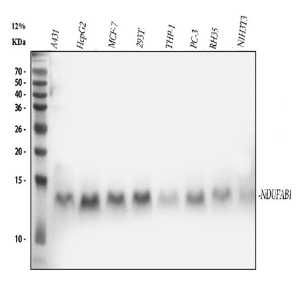
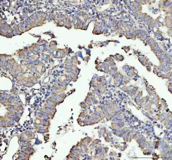
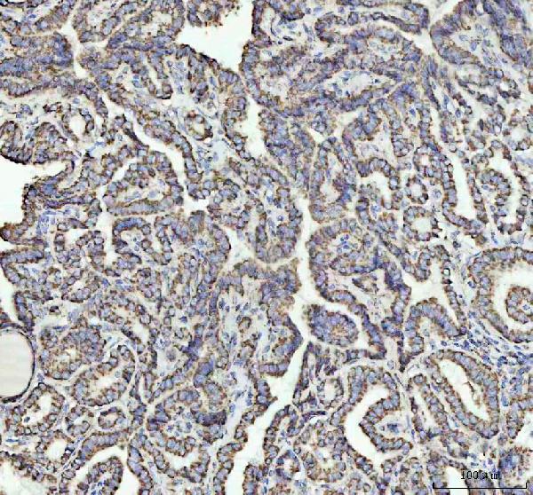
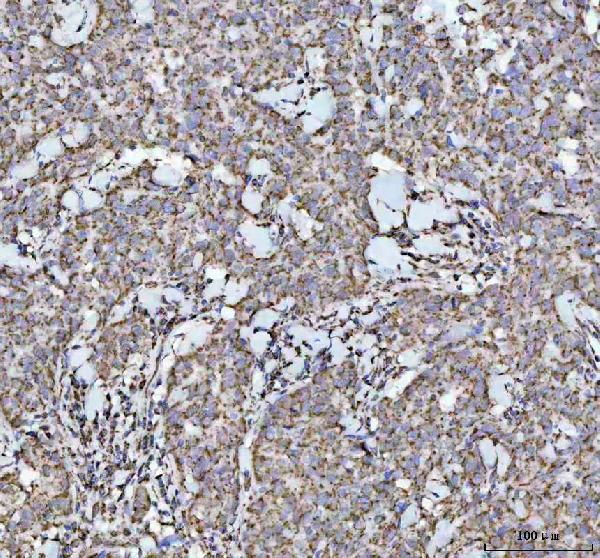
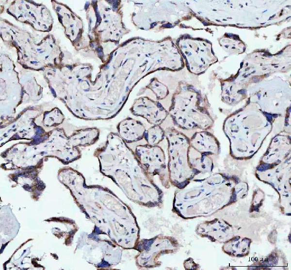
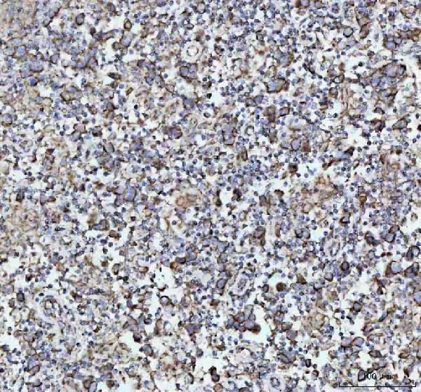
Anti-NDUFAB1 Rabbit Monoclonal Antibody
- SPECIFICATION
- CITATIONS
- PROTOCOLS
- BACKGROUND

Application
| WB, IHC, IP, FC |
|---|---|
| Primary Accession | O14561 |
| Host | Rabbit |
| Isotype | IgG |
| Reactivity | Rat, Human, Mouse |
| Clonality | Monoclonal |
| Format | Liquid |
| Description | Anti-NDUFAB1 Rabbit Monoclonal Antibody . Tested in WB, IHC, IP, Flow Cytometry applications. This antibody reacts with Human, Mouse, Rat. |
| Gene ID | 4706 |
|---|---|
| Other Names | Acyl carrier protein, mitochondrial, ACP, CI-SDAP, NADH-ubiquinone oxidoreductase 9.6 kDa subunit, NDUFAB1 (HGNC:7694) |
| Calculated MW | 10 kDa |
| Application Details | WB 1:500-1:2000 IHC 1:50-1:200 IP 1:50 FC 1:50 |
| Contents | Rabbit IgG in phosphate buffered saline, pH 7.4, 150mM NaCl, 0.02% sodium azide and 50% glycerol, 0.4-0.5mg/ml BSA. |
| Clone Names | Clone: 30N94 |
| Immunogen | A synthesized peptide derived from human NDUFAB1 |
| Purification | Affinity-chromatography |
| Storage | Store at -20°C for one year. For short term storage and frequent use, store at 4°C for up to one month. Avoid repeated freeze-thaw cycles. |
| Name | NDUFAB1 (HGNC:7694) |
|---|---|
| Function | Carrier of the growing fatty acid chain in fatty acid biosynthesis (By similarity) (PubMed:27626371). Accessory and non- catalytic subunit of the mitochondrial membrane respiratory chain NADH dehydrogenase (Complex I), which functions in the transfer of electrons from NADH to the respiratory chain (PubMed:27626371). Accessory protein, of the core iron-sulfur cluster (ISC) assembly complex, that regulates, in association with LYRM4, the stability and the cysteine desulfurase activity of NFS1 and participates in the [2Fe-2S] clusters assembly on the scaffolding protein ISCU (PubMed:31664822). The core iron-sulfur cluster (ISC) assembly complex is involved in the de novo synthesis of a [2Fe-2S] cluster, the first step of the mitochondrial iron-sulfur protein biogenesis. This process is initiated by the cysteine desulfurase complex (NFS1:LYRM4:NDUFAB1) that produces persulfide which is delivered on the scaffold protein ISCU in a FXN- dependent manner. Then this complex is stabilized by FDX2 which provides reducing equivalents to accomplish the [2Fe-2S] cluster assembly. Finally, the [2Fe-2S] cluster is transferred from ISCU to chaperone proteins, including HSCB, HSPA9 and GLRX5 (By similarity). |
| Cellular Location | Mitochondrion |
Research Areas
Citations (0)

Thousands of laboratories across the world have published research that depended on the performance of antibodies from Abcepta to advance their research. Check out links to articles that cite our products in major peer-reviewed journals, organized by research category.
Submit your citation using an Abcepta antibody to
info@abcepta.com, and receive a free "I Love Antibodies" mug.
info@abcepta.com, and receive a free "I Love Antibodies" mug.

Application Protocols
Provided below are standard protocols that you may find useful for product applications.
Abcepta welcomes feedback from its customers.
If you have used an Abcepta product and would like to share how it has performed, please click on the "Submit Review" button and provide the requested information. Our staff will examine and post your review and contact you if needed.
If you have any additional inquiries please email technical services at tech@abcepta.com.
$ 370.00
Cat# ABO16558
Ordering Information
United States
AlbaniaAustraliaAustriaBelgiumBosnia & HerzegovinaBrazilBulgariaCanadaCentral AmericaChinaCroatiaCyprusCzech RepublicDenmarkEstoniaFinlandFranceGermanyGreeceHong KongHungaryIcelandIndiaIndonesiaIrelandIsraelItalyJapanLatviaLithuaniaLuxembourgMacedoniaMalaysiaMaltaNetherlandsNew ZealandNorwayPakistanPolandPortugalRomaniaSerbiaSingaporeSlovakiaSloveniaSouth AfricaSouth KoreaSpainSwedenSwitzerlandTaiwanTurkeyUnited KingdomUnited StatesVietnamWorldwideOthers
USA Headquarters
(888) 735-7227 / (858) 622-0099 or (858) 875-1900
Other Products
Shipping Information
Domestic orders (in stock items)
Shipped out the same day. Orders placed after 1 PM (PST) will ship out the next business day.
International orders
Contact your local distributors
