


 Foundational characteristics of cancer include proliferation, angiogenesis, migration, evasion of apoptosis, and cellular immortality. Find key markers for these cellular processes and antibodies to detect them.
 The SUMOplot™ Analysis Program predicts and scores sumoylation sites in your protein. SUMOylation is a post-translational modification involved in various cellular processes, such as nuclear-cytosolic transport, transcriptional regulation, apoptosis, protein stability, response to stress, and progression through the cell cycle.
 The Autophagy Receptor Motif Plotter predicts and scores autophagy receptor binding sites in your protein. Identifying proteins connected to this pathway is critical to understanding the role of autophagy in physiological as well as pathological processes such as development, differentiation, neurodegenerative diseases, stress, infection, and cancer.
Anti-ICAM1 Antibody Picoband™ (monoclonal, 6F2C3)
- SPECIFICATION
- CITATIONS
- PROTOCOLS
- BACKGROUND

Application
| WB, IHC, FC |
|---|---|
| Primary Accession | P05362 |
| Host | Mouse |
| Isotype | Mouse IgG1 |
| Reactivity | Human |
| Clonality | Monoclonal |
| Format | Lyophilized |
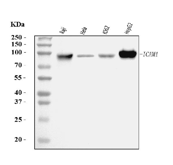
| Description | Anti-ICAM1 Antibody Picoband™ (monoclonal, 6F2C3) . Tested in Flow Cytometry, IHC, WB applications. This antibody reacts with Human. |
| Reconstitution | Adding 0.2 ml of distilled water will yield a concentration of 500 µg/ml. |
| Gene ID | 3383 |
|---|---|
| Other Names | Intercellular adhesion molecule 1, ICAM-1, Major group rhinovirus receptor, CD54, ICAM1 |
| Calculated MW | 90-110 kDa |
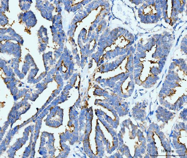
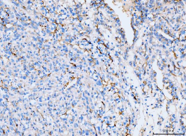
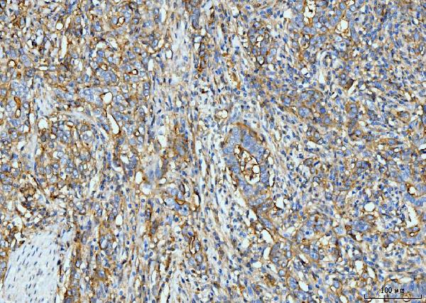
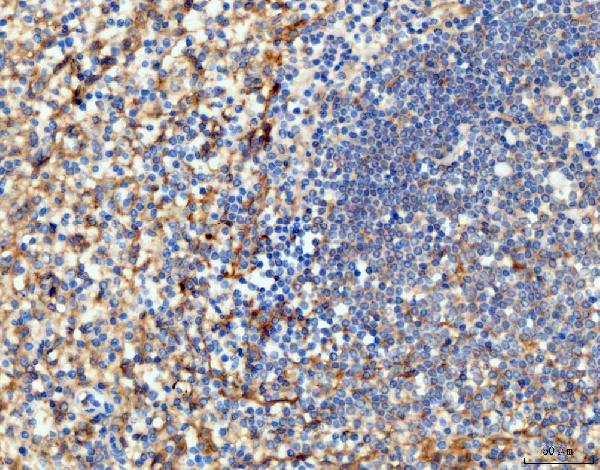
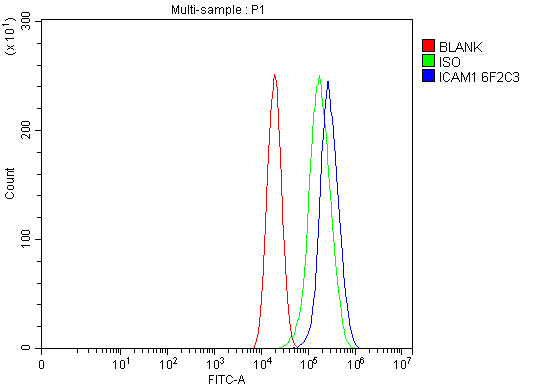
| Application Details | Western blot, 0.25-0.5 µg/ml, Human Immunohistochemistry(Paraffin-embedded Section), 2-5 µg/ml, Human Flow Cytometry, 1-3 µg/1x10^6 cells, Human |
| Contents | Each vial contains 4 mg Trehalose, 0.9 mg NaCl and 0.2 mg Na2HPO4. |
| Clone Names | Clone: 6F2C3 |
| Immunogen | E. coli-derived human ICAM1 recombinant protein (Position: Q28-R268). |
| Purification | Immunogen affinity purified. |
| Storage | At -20°C for one year from date of receipt. After reconstitution, at 4°C for one month. It can also be aliquotted and stored frozen at -20°C for six months. Avoid repeated freezing and thawing. |
| Name | ICAM1 |
|---|---|
| Function | ICAM proteins are ligands for the leukocyte adhesion protein LFA-1 (integrin alpha-L/beta-2). During leukocyte trans-endothelial migration, ICAM1 engagement promotes the assembly of endothelial apical cups through ARHGEF26/SGEF and RHOG activation. |
| Cellular Location | Membrane; Single-pass type I membrane protein. |

Thousands of laboratories across the world have published research that depended on the performance of antibodies from Abcepta to advance their research. Check out links to articles that cite our products in major peer-reviewed journals, organized by research category.
info@abcepta.com, and receive a free "I Love Antibodies" mug.

Provided below are standard protocols that you may find useful for product applications.
Background
CD54, also known as ICAM-1. Intercellular adhesion molecule-1 (ICAM1) is a ligand for lymphocyte function-associated (LFA) antigens. ICAM-1 is an integral membrane protein, a member of the immunoglobulin superfamily, and a ligand for LFA-1, a beta 2 leukocyte integrin. This protein is the major human rhinovirus receptor. The ICAM1 gene is mapped to human chromosome 19. In humans, lymphocyte adhesion to cells is mediated by the protein heterodimer CD11a/CD18 (Leu-CAMa, LFA-1) and its ligand CD54 (ICAM-1).
If you have used an Abcepta product and would like to share how it has performed, please click on the "Submit Review" button and provide the requested information. Our staff will examine and post your review and contact you if needed.
If you have any additional inquiries please email technical services at tech@abcepta.com.
Ordering Information
Other Products
Shipping Information
