


 Foundational characteristics of cancer include proliferation, angiogenesis, migration, evasion of apoptosis, and cellular immortality. Find key markers for these cellular processes and antibodies to detect them.
 The SUMOplot™ Analysis Program predicts and scores sumoylation sites in your protein. SUMOylation is a post-translational modification involved in various cellular processes, such as nuclear-cytosolic transport, transcriptional regulation, apoptosis, protein stability, response to stress, and progression through the cell cycle.
 The Autophagy Receptor Motif Plotter predicts and scores autophagy receptor binding sites in your protein. Identifying proteins connected to this pathway is critical to understanding the role of autophagy in physiological as well as pathological processes such as development, differentiation, neurodegenerative diseases, stress, infection, and cancer.
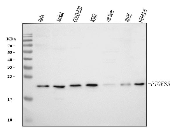
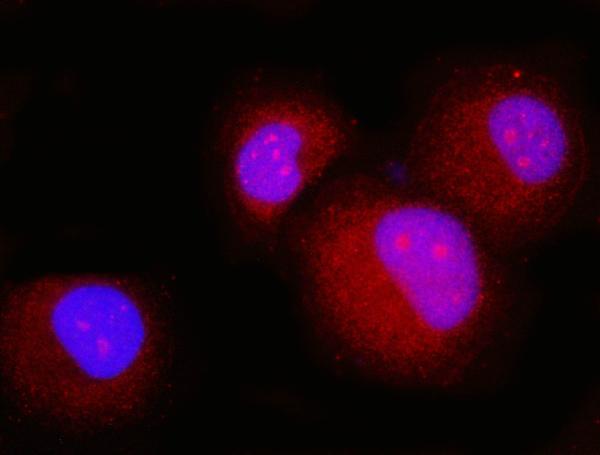
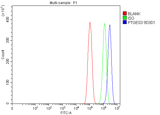
Anti-p23/PTGES3 Antibody Picoband™ (monoclonal, 9D3D1)
- SPECIFICATION
- CITATIONS
- PROTOCOLS
- BACKGROUND

Application
| WB, IF, ICC, FC |
|---|---|
| Primary Accession | Q15185 |
| Host | Mouse |
| Isotype | Mouse IgG2b |
| Reactivity | Rat, Human, Mouse |
| Clonality | Monoclonal |
| Format | Lyophilized |
| Description | Anti-p23/PTGES3 Antibody Picoband™ (monoclonal, 9D3D1) . Tested in Flow Cytometry, IF, ICC, WB applications. This antibody reacts with Human, Mouse, Rat. |
| Reconstitution | Adding 0.2 ml of distilled water will yield a concentration of 500 µg/ml. |
| Gene ID | 10728 |
|---|---|
| Other Names | Prostaglandin E synthase 3, 5.3.99.3, Cytosolic prostaglandin E2 synthase, cPGES, Hsp90 co-chaperone, Progesterone receptor complex p23, Telomerase-binding protein p23, PTGES3, P23, TEBP |
| Calculated MW | 23 kDa |
| Application Details | Western blot, 0.25-0.5 µg/ml, Human, Mouse, Rat Immunocytochemistry/Immunofluorescence, 5 µg/ml, Human Flow Cytometry, 1-3 µg/1x10^6 cells, Human |
| Contents | Each vial contains 4 mg Trehalose, 0.9 mg NaCl and 0.2 mg Na2HPO4. |
| Clone Names | Clone: 9D3D1 |
| Immunogen | E.coli-derived human p23/PTGES3 recombinant protein (Position: M1-K79). |
| Purification | Immunogen affinity purified. |
| Storage | At -20°C for one year from date of receipt. After reconstitution, at 4°C for one month. It can also be aliquotted and stored frozen at -20°C for six months. Avoid repeated freezing and thawing. |
| Name | PTGES3 |
|---|---|
| Synonyms | P23, TEBP |
| Function | Cytosolic prostaglandin synthase that catalyzes the oxidoreduction of prostaglandin endoperoxide H2 (PGH2) to prostaglandin E2 (PGE2) (PubMed:10922363). Molecular chaperone that localizes to genomic response elements in a hormone-dependent manner and disrupts receptor-mediated transcriptional activation, by promoting disassembly of transcriptional regulatory complexes (PubMed:11274138, PubMed:12077419). Facilitates HIF alpha proteins hydroxylation via interaction with EGLN1/PHD2, leading to recruit EGLN1/PHD2 to the HSP90 pathway (PubMed:24711448). |
| Cellular Location | Cytoplasm {ECO:0000250|UniProtKB:Q3ZBF7}. |

Thousands of laboratories across the world have published research that depended on the performance of antibodies from Abcepta to advance their research. Check out links to articles that cite our products in major peer-reviewed journals, organized by research category.
info@abcepta.com, and receive a free "I Love Antibodies" mug.

Provided below are standard protocols that you may find useful for product applications.
Background
Prostaglandin E synthase 3 (cytosolic)is anenzymethat in humans is encoded by the PTGES3gene. It is mapped to 12q13.3; 12. This gene encodes an enzyme that converts prostaglandin endoperoxide H2 (PGH2) to prostaglandin E2 (PGE2). This protein functions as a co-chaperone with heat shock protein 90 (HSP90), localizing to response elements in DNA and disrupting transcriptional activation complexes. Alternative splicing results in multiple transcript variants. There are multiple pseudogenes of this gene on several different chromosomes.
If you have used an Abcepta product and would like to share how it has performed, please click on the "Submit Review" button and provide the requested information. Our staff will examine and post your review and contact you if needed.
If you have any additional inquiries please email technical services at tech@abcepta.com.
Ordering Information
Other Products
Shipping Information
