


 Foundational characteristics of cancer include proliferation, angiogenesis, migration, evasion of apoptosis, and cellular immortality. Find key markers for these cellular processes and antibodies to detect them.
 The SUMOplot™ Analysis Program predicts and scores sumoylation sites in your protein. SUMOylation is a post-translational modification involved in various cellular processes, such as nuclear-cytosolic transport, transcriptional regulation, apoptosis, protein stability, response to stress, and progression through the cell cycle.
 The Autophagy Receptor Motif Plotter predicts and scores autophagy receptor binding sites in your protein. Identifying proteins connected to this pathway is critical to understanding the role of autophagy in physiological as well as pathological processes such as development, differentiation, neurodegenerative diseases, stress, infection, and cancer.
> home > Products > Primary Antibodies > Microbiology > Anti-Beclin 1 Antibody Picoband™ (monoclonal, 2D12A3)
Anti-Beclin 1 Antibody Picoband™ (monoclonal, 2D12A3)
- SPECIFICATION
- CITATIONS
- PROTOCOLS
- BACKGROUND

Application
| WB, IHC, IF, ICC, FC |
|---|---|
| Primary Accession | Q14457 |
| Host | Mouse |
| Isotype | Mouse IgG1 |
| Reactivity | Rat, Human, Mouse |
| Clonality | Monoclonal |
| Format | Lyophilized |
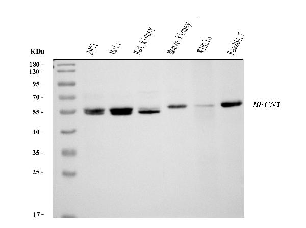
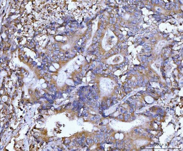
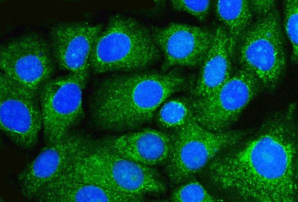
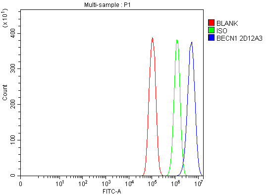
| Description | Anti-Beclin 1 Antibody Picoband™ (monoclonal, 2D12A3) . Tested in Flow Cytometry, IF, IHC, ICC, WB applications. This antibody reacts with Human, Mouse, Rat. |
| Reconstitution | Adding 0.2 ml of distilled water will yield a concentration of 500 µg/ml. |
| Gene ID | 8678 |
|---|---|
| Other Names | Beclin-1, Coiled-coil myosin-like BCL2-interacting protein, Protein GT197, Beclin-1-C 35 kDa, Beclin-1-C 37 kDa, BECN1, GT197 |
| Calculated MW | 52-60 kDa |
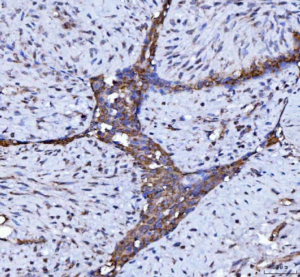
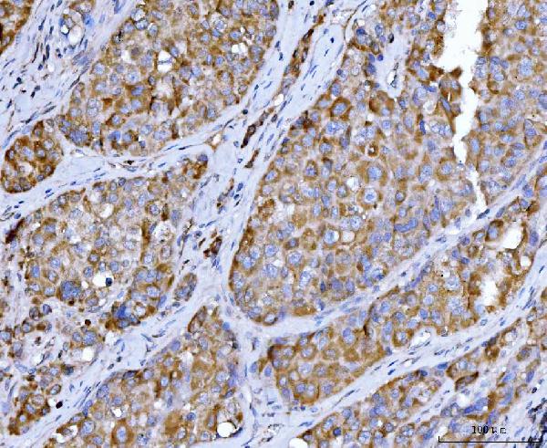
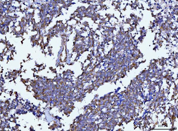
| Application Details | Western blot, 0.25-0.5 µg/ml, Human, Mouse, Rat Immunohistochemistry(Paraffin-embedded Section), 2-5 µg/ml, Human Immunocytochemistry/Immunofluorescence, 5 µg/ml, Human Flow Cytometry, 1-3 µg/1x10^6 cells, Human |
| Contents | Each vial contains 4 mg Trehalose, 0.9 mg NaCl and 0.2 mg Na2HPO4. |
| Clone Names | Clone: 2D12A3 |
| Immunogen | E.coli-derived human Beclin 1 recombinant protein (Position: M1-S354). Human Beclin 1 shares 97% amino acid (aa) sequence identity with both mouse and rat Beclin 1. |
| Purification | Immunogen affinity purified. |
| Storage | At -20°C for one year from date of receipt. After reconstitution, at 4°C for one month. It can also be aliquotted and stored frozen at -20°C for six months. Avoid repeated freezing and thawing. |
| Name | BECN1 |
|---|---|
| Synonyms | GT197 |
| Function | Plays a central role in autophagy (PubMed:18570871, PubMed:21358617, PubMed:23184933, PubMed:23974797, PubMed:25484083, PubMed:28445460, PubMed:37776275). Acts as a core subunit of the PI3K complex that mediates formation of phosphatidylinositol 3-phosphate; different complex forms are believed to play a role in multiple membrane trafficking pathways: PI3KC3-C1 is involved in initiation of autophagosomes and PI3KC3-C2 in maturation of autophagosomes and endocytosis. Involved in regulation of degradative endocytic trafficking and required for the abscission step in cytokinesis, probably in the context of PI3KC3-C2 (PubMed:20208530, PubMed:20643123, PubMed:23974797, PubMed:26783301). Essential for the formation of PI3KC3-C2 but not PI3KC3-C1 PI3K complex forms. Involved in endocytosis (PubMed:25275521). May play a role in antiviral host defense. |
| Cellular Location | Cytoplasm. Golgi apparatus, trans-Golgi network membrane; Peripheral membrane protein. Endosome membrane; Peripheral membrane protein. Endoplasmic reticulum membrane; Peripheral membrane protein. Mitochondrion membrane; Peripheral membrane protein. Endosome {ECO:0000250|UniProtKB:O88597} Cytoplasmic vesicle, autophagosome. Note=Interaction with ATG14 promotes translocation to autophagosomes. Expressed in dendrites and cell bodies of cerebellar Purkinje cells (By similarity) {ECO:0000250|UniProtKB:O88597, ECO:0000269|PubMed:19050071} [Beclin-1-C 37 kDa]: Mitochondrion {ECO:0000250|UniProtKB:O88597} |
| Tissue Location | Ubiquitous. |
Research Areas

Abcepta welcomes feedback from its customers.
If you have used an Abcepta product and would like to share how it has performed, please click on the "Submit Review" button and provide the requested information. Our staff will examine and post your review and contact you if needed.
If you have any additional inquiries please email technical services at tech@abcepta.com.
$ 370.00
Cat# ABO16255
Ordering Information
United States
AlbaniaAustraliaAustriaBelgiumBosnia & HerzegovinaBrazilBulgariaCanadaCentral AmericaChinaCroatiaCyprusCzech RepublicDenmarkEstoniaFinlandFranceGermanyGreeceHong KongHungaryIcelandIndiaIndonesiaIrelandIsraelItalyJapanLatviaLithuaniaLuxembourgMacedoniaMalaysiaMaltaNetherlandsNew ZealandNorwayPakistanPolandPortugalRomaniaSerbiaSingaporeSlovakiaSloveniaSouth AfricaSouth KoreaSpainSwedenSwitzerlandTaiwanTurkeyUnited KingdomUnited StatesVietnamWorldwideOthersMexico
USA Headquarters
(888) 735-7227 / (858) 622-0099 or (858) 875-1900
Other Products
Shipping Information
Domestic orders (in stock items)
Shipped out the same day. Orders placed after 1 PM (PST) will ship out the next business day.
International orders
Contact your local distributors
