


 Foundational characteristics of cancer include proliferation, angiogenesis, migration, evasion of apoptosis, and cellular immortality. Find key markers for these cellular processes and antibodies to detect them.
 The SUMOplot™ Analysis Program predicts and scores sumoylation sites in your protein. SUMOylation is a post-translational modification involved in various cellular processes, such as nuclear-cytosolic transport, transcriptional regulation, apoptosis, protein stability, response to stress, and progression through the cell cycle.
 The Autophagy Receptor Motif Plotter predicts and scores autophagy receptor binding sites in your protein. Identifying proteins connected to this pathway is critical to understanding the role of autophagy in physiological as well as pathological processes such as development, differentiation, neurodegenerative diseases, stress, infection, and cancer.
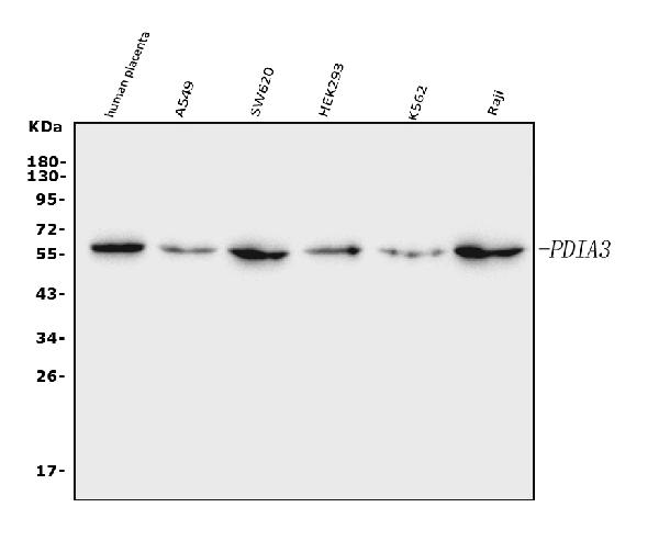
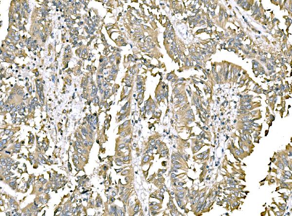
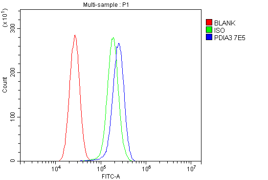
Anti-ERp57 Antibody Picoband™ (monoclonal, 7E5)
- SPECIFICATION
- CITATIONS
- PROTOCOLS
- BACKGROUND

Application
| WB, IHC, FC |
|---|---|
| Primary Accession | P30101 |
| Host | Mouse |
| Isotype | Mouse IgG1 |
| Reactivity | Human |
| Clonality | Monoclonal |
| Format | Lyophilized |
| Description | Anti-ERp57 Antibody Picoband™ (monoclonal, 7E5) . Tested in Flow Cytometry, IHC, WB applications. This antibody reacts with Human. |
| Reconstitution | Adding 0.2 ml of distilled water will yield a concentration of 500 µg/ml. |
| Gene ID | 2923 |
|---|---|
| Other Names | Protein disulfide-isomerase A3, 5.3.4.1, 58 kDa glucose-regulated protein, 58 kDa microsomal protein, p58, Disulfide isomerase ER-60, Endoplasmic reticulum resident protein 57, ER protein 57, ERp57, Endoplasmic reticulum resident protein 60, ER protein 60, ERp60, PDIA3 (HGNC:4606), ERP57, ERP60, GRP58 |
| Calculated MW | 57 kDa |
| Application Details | Western blot, 0.25-0.5 µg/ml, Human Immunohistochemistry(Paraffin-embedded Section), 2-5 µg/ml, Human Flow Cytometry, 1-3 µg/1x10^6 cells, Human |
| Contents | Each vial contains 4 mg Trehalose, 0.9 mg NaCl and 0.2 mg Na2HPO4. |
| Clone Names | Clone: 7E5 |
| Immunogen | A synthetic peptide corresponding to a sequence at the C-terminus of human ERp57, different from the related mouse and rat sequences by two amino acids. |
| Purification | Immunogen affinity purified. |
| Storage | At -20°C for one year from date of receipt. After reconstitution, at 4°C for one month. It can also be aliquotted and stored frozen at -20°C for six months. Avoid repeated freezing and thawing. |
| Name | PDIA3 (HGNC:4606) |
|---|---|
| Synonyms | ERP57, ERP60, GRP58 |
| Function | Protein disulfide isomerase that catalyzes the formation, isomerization, and reduction or oxidation of disulfide bonds in client proteins and functions as a protein folding chaperone (PubMed:11825568, PubMed:16193070, PubMed:27897272, PubMed:36104323, PubMed:7487104). Core component of the major histocompatibility complex class I (MHC I) peptide loading complex where it functions as an essential folding chaperone for TAPBP. Through TAPBP, assists the dynamic assembly of the MHC I complex with high affinity antigens in the endoplasmic reticulum. Therefore, plays a crucial role in the presentation of antigens to cytotoxic T cells in adaptive immunity (PubMed:35948544, PubMed:36104323). |
| Cellular Location | Endoplasmic reticulum. Endoplasmic reticulum lumen {ECO:0000250|UniProtKB:P11598}. Melanosome Note=Identified by mass spectrometry in melanosome fractions from stage I to stage IV (PubMed:12643545). |
| Tissue Location | Detected in the flagellum and head region of spermatozoa (at protein level) (PubMed:20400973). Expressed in liver, stomach and colon (at protein level). Expressed in gastric parietal cells and chief cells (at protein level) (PubMed:24188822) |

Thousands of laboratories across the world have published research that depended on the performance of antibodies from Abcepta to advance their research. Check out links to articles that cite our products in major peer-reviewed journals, organized by research category.
info@abcepta.com, and receive a free "I Love Antibodies" mug.

Provided below are standard protocols that you may find useful for product applications.
Background
PDIA3 (Protein disulfide isomerase family A, member 3), also called GRP58, Erp57 or ER60, is an isomerase enzyme. It is mapped on 15q15.3. PDIA3 is also part of the major histocompatibility complex (MHC) class I peptide-loading complex, which is essential for formation of the final antigen conformation and export from the endoplasmic reticulum to the cell surface. This gene encodes a protein of the endoplasmic reticulum that interacts with lectin chaperones calreticulin and calnexin to modulate folding of newly synthesized glycoproteins. The protein was once thought to be a phospholipase; however, it has been demonstrated that the protein actually has protein disulfide isomerase activity. It is thought that complexes of lectins and this protein mediate protein folding by promoting formation of disulfide bonds in their glycoprotein substrates.
If you have used an Abcepta product and would like to share how it has performed, please click on the "Submit Review" button and provide the requested information. Our staff will examine and post your review and contact you if needed.
If you have any additional inquiries please email technical services at tech@abcepta.com.
Ordering Information
Other Products
Shipping Information
