


 Foundational characteristics of cancer include proliferation, angiogenesis, migration, evasion of apoptosis, and cellular immortality. Find key markers for these cellular processes and antibodies to detect them.
 The SUMOplot™ Analysis Program predicts and scores sumoylation sites in your protein. SUMOylation is a post-translational modification involved in various cellular processes, such as nuclear-cytosolic transport, transcriptional regulation, apoptosis, protein stability, response to stress, and progression through the cell cycle.
 The Autophagy Receptor Motif Plotter predicts and scores autophagy receptor binding sites in your protein. Identifying proteins connected to this pathway is critical to understanding the role of autophagy in physiological as well as pathological processes such as development, differentiation, neurodegenerative diseases, stress, infection, and cancer.
Anti-PDIA6 Antibody Picoband™ (monoclonal, 3H5E7)
- SPECIFICATION
- CITATIONS
- PROTOCOLS
- BACKGROUND

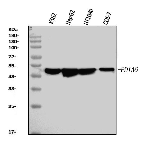
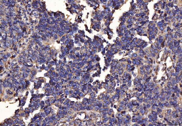
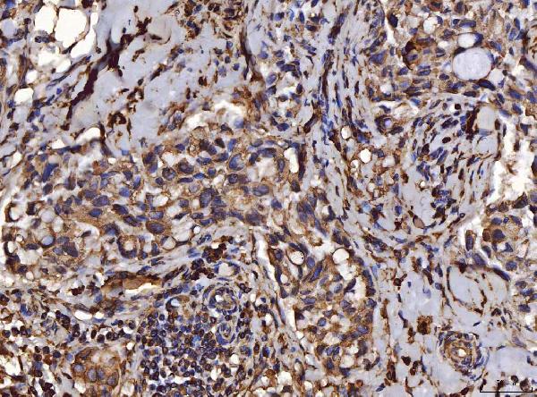
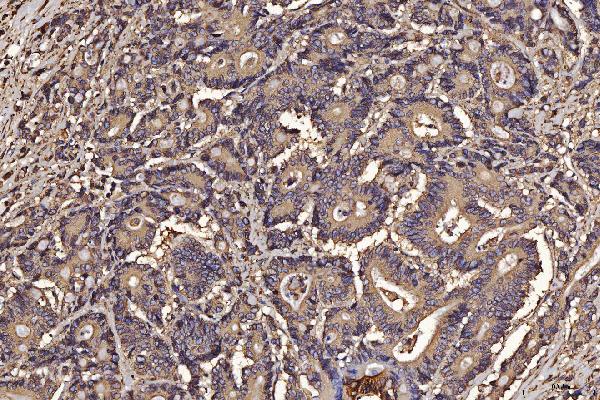
Application
| WB, IHC, IF, ICC, FC |
|---|---|
| Primary Accession | Q15084 |
| Host | Mouse |
| Isotype | Mouse IgG2b |
| Reactivity | Human, Monkey |
| Clonality | Monoclonal |
| Format | Lyophilized |
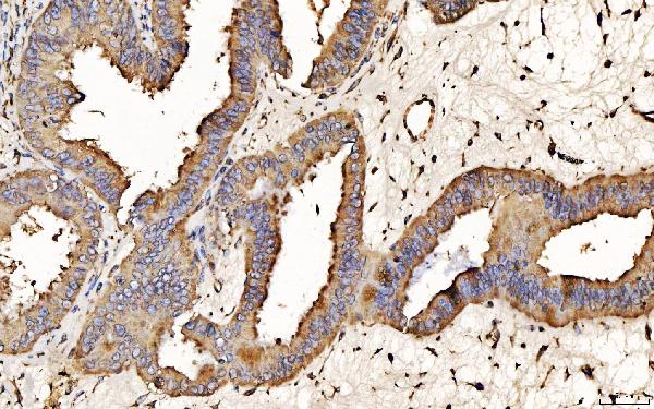
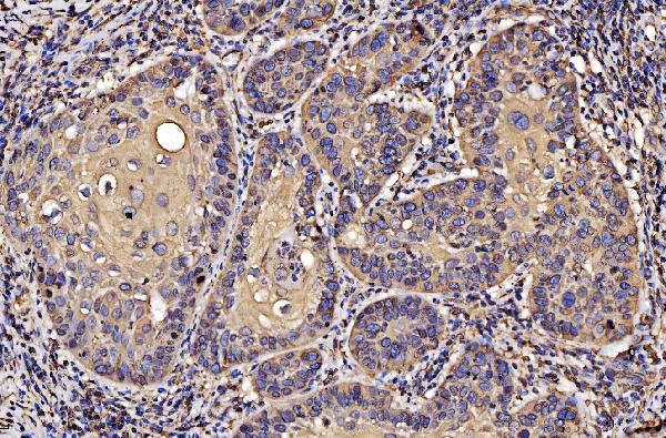
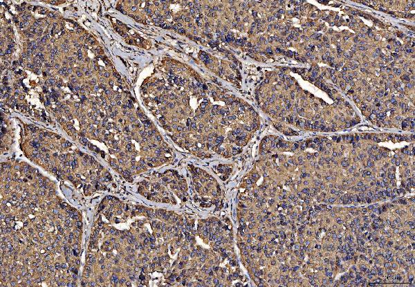
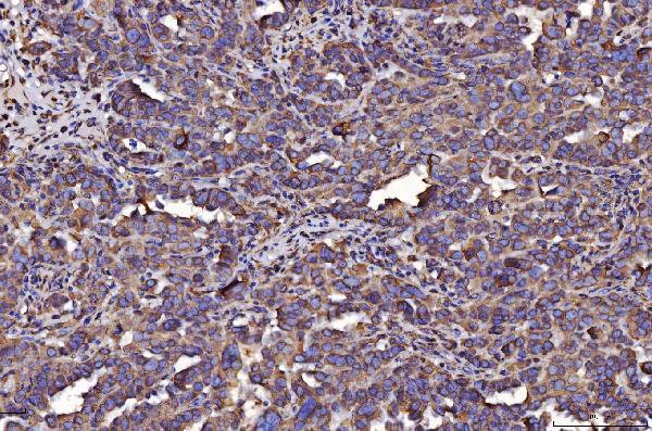
| Description | Anti-PDIA6 Antibody Picoband™ (monoclonal, 3H5E7) . Tested in Flow Cytometry, IF, IHC, ICC, WB applications. This antibody reacts with Human, Monkey. |
| Reconstitution | Adding 0.2 ml of distilled water will yield a concentration of 500 µg/ml. |
| Gene ID | 10130 |
|---|---|
| Other Names | Protein disulfide-isomerase A6, 5.3.4.1, Endoplasmic reticulum protein 5, ER protein 5, ERp5, Protein disulfide isomerase P5, Thioredoxin domain-containing protein 7, PDIA6, ERP5, P5, TXNDC7 |
| Calculated MW | 48 kDa |
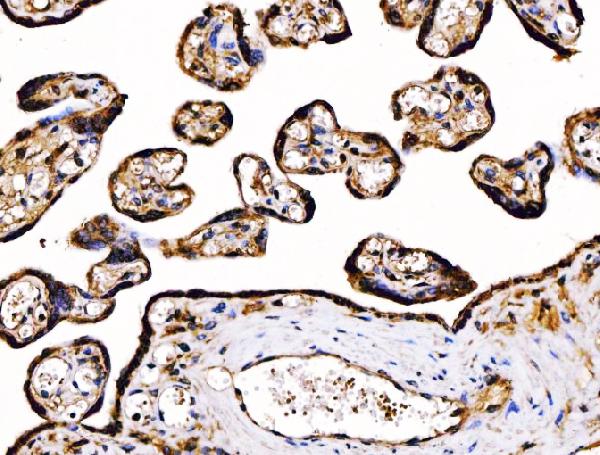
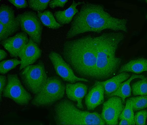
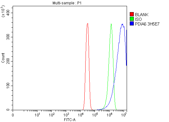
| Application Details | Western blot, 0.25-0.5 µg/ml, Human, Monkey Immunohistochemistry(Paraffin-embedded Section), 2-5 µg/ml, Human Immunocytochemistry/Immunofluorescence, 5 µg/ml, Human Flow Cytometry, 1-3 µg/1x10^6 cells, Human |
| Contents | Each vial contains 4 mg Trehalose, 0.9 mg NaCl and 0.2 mg Na2HPO4. |
| Clone Names | Clone: 3H5E7 |
| Immunogen | E.coli-derived human PDIA6 recombinant protein (Position: L20-L440). |
| Purification | Immunogen affinity purified. |
| Storage | At -20°C for one year from date of receipt. After reconstitution, at 4°C for one month. It can also be aliquotted and stored frozen at -20°C for six months. Avoid repeated freezing and thawing. |
| Name | PDIA6 |
|---|---|
| Synonyms | ERP5, P5, TXNDC7 |
| Function | May function as a chaperone that inhibits aggregation of misfolded proteins (PubMed:12204115). Negatively regulates the unfolded protein response (UPR) through binding to UPR sensors such as ERN1, which in turn inactivates ERN1 signaling (PubMed:24508390). May also regulate the UPR via the EIF2AK3 UPR sensor (PubMed:24508390). Plays a role in platelet aggregation and activation by agonists such as convulxin, collagen and thrombin (PubMed:15466936). |
| Cellular Location | Endoplasmic reticulum lumen. Cell membrane. Melanosome. Note=Identified by mass spectrometry in melanosome fractions from stage I to stage IV (PubMed:12643545) |
| Tissue Location | Expressed in platelets (at protein level). |

Thousands of laboratories across the world have published research that depended on the performance of antibodies from Abcepta to advance their research. Check out links to articles that cite our products in major peer-reviewed journals, organized by research category.
info@abcepta.com, and receive a free "I Love Antibodies" mug.

Provided below are standard protocols that you may find useful for product applications.
Background
This gene encodes a member of the disulfide isomerase (PDI) family of endoplasmic reticulum (ER) proteins that catalyze protein folding and thiol-disulfide interchange reactions. The encoded protein has an N-terminal ER-signal sequence, two catalytically active thioredoxin (TRX) domains, a TRX-like domain, and a C-terminal ER-retention sequence. This protein inhibits the aggregation of misfolded proteins and exhibits both isomerase and chaperone activity. Alternative splicing results in multiple transcript variants encoding different isoforms.
If you have used an Abcepta product and would like to share how it has performed, please click on the "Submit Review" button and provide the requested information. Our staff will examine and post your review and contact you if needed.
If you have any additional inquiries please email technical services at tech@abcepta.com.
Ordering Information
Other Products
Shipping Information
