


 Foundational characteristics of cancer include proliferation, angiogenesis, migration, evasion of apoptosis, and cellular immortality. Find key markers for these cellular processes and antibodies to detect them.
 The SUMOplot™ Analysis Program predicts and scores sumoylation sites in your protein. SUMOylation is a post-translational modification involved in various cellular processes, such as nuclear-cytosolic transport, transcriptional regulation, apoptosis, protein stability, response to stress, and progression through the cell cycle.
 The Autophagy Receptor Motif Plotter predicts and scores autophagy receptor binding sites in your protein. Identifying proteins connected to this pathway is critical to understanding the role of autophagy in physiological as well as pathological processes such as development, differentiation, neurodegenerative diseases, stress, infection, and cancer.
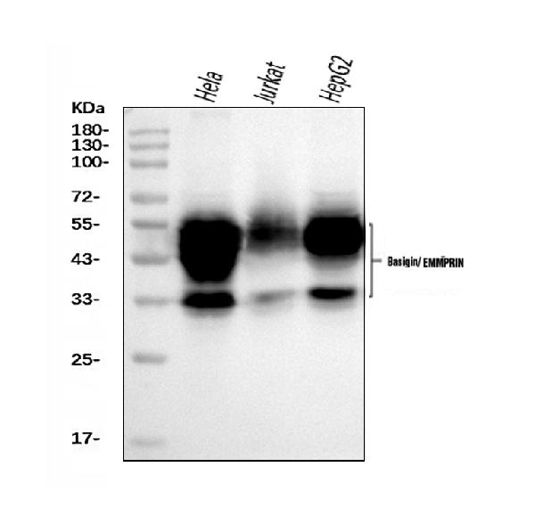
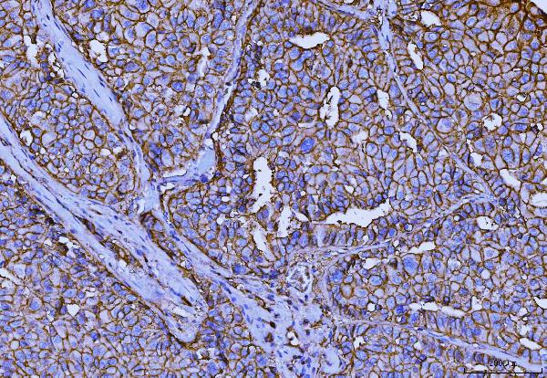
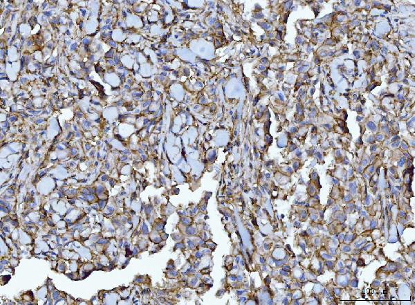
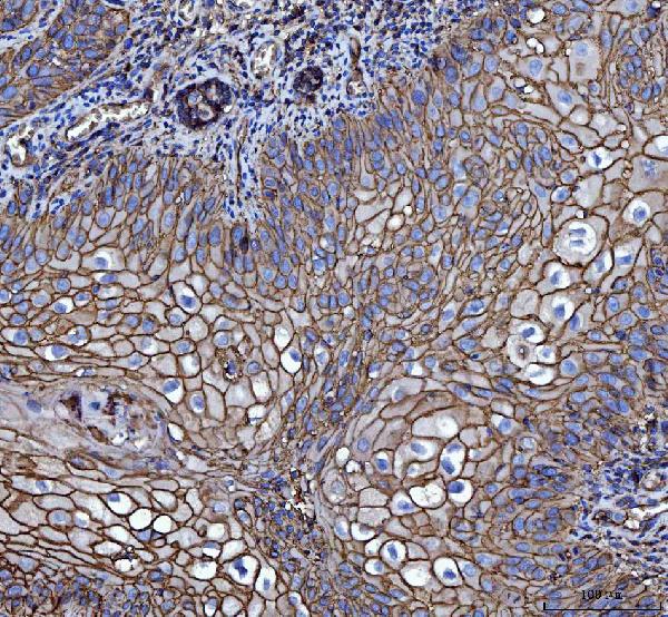
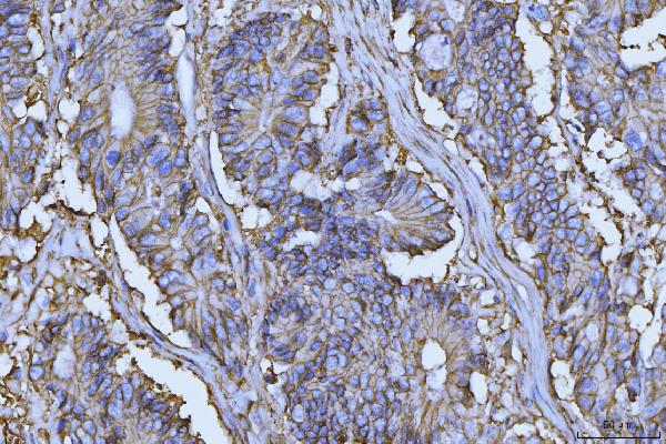
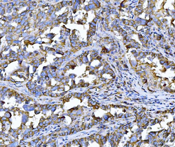
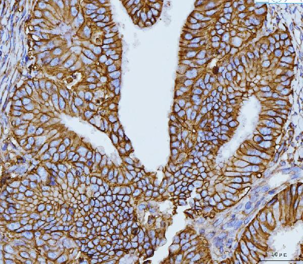
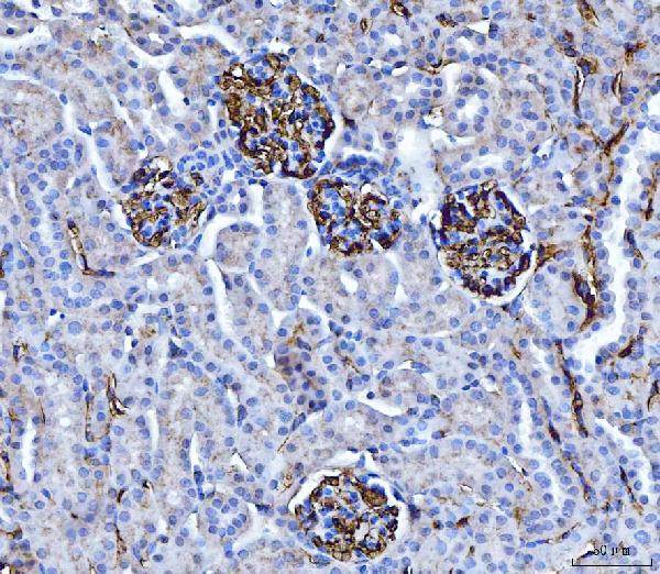
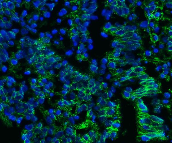
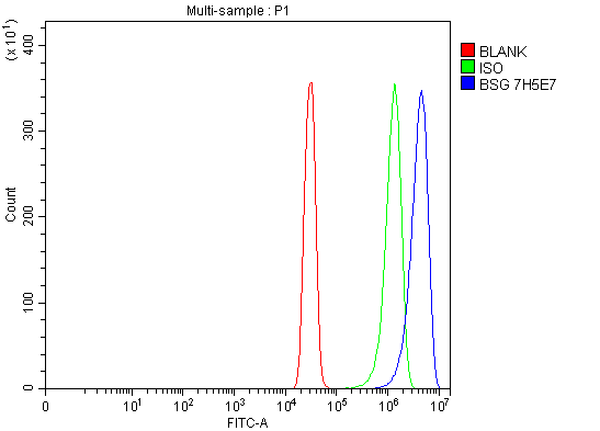
Anti-CD147/Emmprin Antibody Picoband™ (monoclonal, 7H5E7)
- SPECIFICATION
- CITATIONS
- PROTOCOLS
- BACKGROUND

Application
| WB, IHC, IF, FC |
|---|---|
| Primary Accession | P35613 |
| Host | Mouse |
| Isotype | Mouse IgG2a |
| Reactivity | Human, Mouse |
| Clonality | Monoclonal |
| Format | Lyophilized |
| Description | Anti-CD147/Emmprin Antibody Picoband™ (monoclonal, 7H5E7) . Tested in Flow Cytometry, IF, IHC, WB applications. This antibody reacts with Human, Mouse. |
| Reconstitution | Adding 0.2 ml of distilled water will yield a concentration of 500 µg/ml. |
| Gene ID | 682 |
|---|---|
| Other Names | Basigin, 5F7, Collagenase stimulatory factor, Extracellular matrix metalloproteinase inducer, EMMPRIN, Hepatoma-associated antigen, HAb18G, Leukocyte activation antigen M6, OK blood group antigen, Tumor cell-derived collagenase stimulatory factor, TCSF, CD147, BSG (HGNC:1116) |
| Calculated MW | 35-60 kDa |
| Application Details | Western blot, 0.25-0.5 µg/ml, Human Immunohistochemistry(Paraffin-embedded Section), 2-5 µg/ml, Human, Mouse Immunocytochemistry, 5 µg/ml, Human Flow Cytometry, 1-3 µg/1x10^6 cells, Human |
| Contents | Each vial contains 4 mg Trehalose, 0.9 mg NaCl and 0.2 mg Na2HPO4. |
| Clone Names | Clone: 7H5E7 |
| Immunogen | E.coli-derived human CD147/Emmprin recombinant protein (Position: E138-A323). Human CD147/Emmprin shares 51.1% and 51.9% amino acid (aa) sequence identity with mouse and rat CD147/Emmprin, respectively. |
| Purification | Immunogen affinity purified. |
| Storage | At -20°C for one year from date of receipt. After reconstitution, at 4°C for one month. It can also be aliquotted and stored frozen at -20°C for six months. Avoid repeated freezing and thawing. |
| Name | BSG (HGNC:1116) |
|---|---|
| Function | [Isoform 1]: Essential for normal retinal maturation and development (By similarity). Acts as a retinal cell surface receptor for NXNL1 and plays an important role in NXNL1-mediated survival of retinal cone photoreceptors (PubMed:25957687). In association with glucose transporter SLC16A1/GLUT1 and NXNL1, promotes retinal cone survival by enhancing aerobic glycolysis and accelerating the entry of glucose into photoreceptors (PubMed:25957687). May act as a potent stimulator of IL6 secretion in multiple cell lines that include monocytes (PubMed:21620857). |
| Cellular Location | Melanosome. Note=Identified by mass spectrometry in melanosome fractions from stage I to stage IV. [Isoform 2]: Cell membrane; Single-pass type I membrane protein {ECO:0000250|UniProtKB:P26453}. Endosome Endoplasmic reticulum membrane; Single- pass type I membrane protein {ECO:0000250|UniProtKB:P26453} Basolateral cell membrane; Single-pass type I membrane protein {ECO:0000250|UniProtKB:P26453} [Isoform 4]: Cell membrane; Single-pass type I membrane protein {ECO:0000250|UniProtKB:P26453} |
| Tissue Location | [Isoform 1]: Retina-specific (PubMed:25957687). Expressed in retinal cone photoreceptors (at protein level) (PubMed:25957687). [Isoform 3]: Highly expressed in the bone marrow, fetal liver, lung, testis and thymus. |

Thousands of laboratories across the world have published research that depended on the performance of antibodies from Abcepta to advance their research. Check out links to articles that cite our products in major peer-reviewed journals, organized by research category.
info@abcepta.com, and receive a free "I Love Antibodies" mug.

Provided below are standard protocols that you may find useful for product applications.
Background
Emmprin, extracellular matrix metalloproteinase inducer, also known as Emmprin (BSG) or cluster of differentiation 147 (CD147) is a protein that in humans is encoded by the Emmprin gene. The human BSG gene is mapped to 19p13.3. This protein is a determinant for the Ok blood group system. BSG has been shown to be an essential receptor on red blood cells for the malaria parasite. It is a member of the immunoglobulin superfamily, with a structure related to the putative primordial form of the family. As members of the immunoglobulin superfamily, it plays fundamental roles in intercellular recognition involved in various immunologic phenomena, differentiation, and development. BSG is thought also to play a role in intercellular recognition. It also regulates several distinct functions, such as spermatogenesis, expression of the monocarboxylate transporter and the responsiveness of lymphocytes. BSG is a type I integral membrane receptor that has many ligands, including the cyclophilin (CyP) proteins Cyp-A and CyP-B and certain integrins. It is expressed by many cell types, including epithelial cells, endothelial cells and leukocytes.
If you have used an Abcepta product and would like to share how it has performed, please click on the "Submit Review" button and provide the requested information. Our staff will examine and post your review and contact you if needed.
If you have any additional inquiries please email technical services at tech@abcepta.com.
Ordering Information
Other Products
Shipping Information
