


 Foundational characteristics of cancer include proliferation, angiogenesis, migration, evasion of apoptosis, and cellular immortality. Find key markers for these cellular processes and antibodies to detect them.
 The SUMOplot™ Analysis Program predicts and scores sumoylation sites in your protein. SUMOylation is a post-translational modification involved in various cellular processes, such as nuclear-cytosolic transport, transcriptional regulation, apoptosis, protein stability, response to stress, and progression through the cell cycle.
 The Autophagy Receptor Motif Plotter predicts and scores autophagy receptor binding sites in your protein. Identifying proteins connected to this pathway is critical to understanding the role of autophagy in physiological as well as pathological processes such as development, differentiation, neurodegenerative diseases, stress, infection, and cancer.
Anti-Cdk2 Antibody Picoband™ (monoclonal, 6D5B5)
- SPECIFICATION
- CITATIONS
- PROTOCOLS
- BACKGROUND

Application
| WB, IHC |
|---|---|
| Primary Accession | P24941 |
| Host | Mouse |
| Isotype | Mouse IgG2b |
| Reactivity | Rat, Human, Mouse |
| Clonality | Monoclonal |
| Format | Lyophilized |
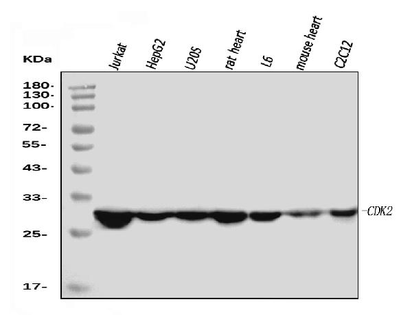
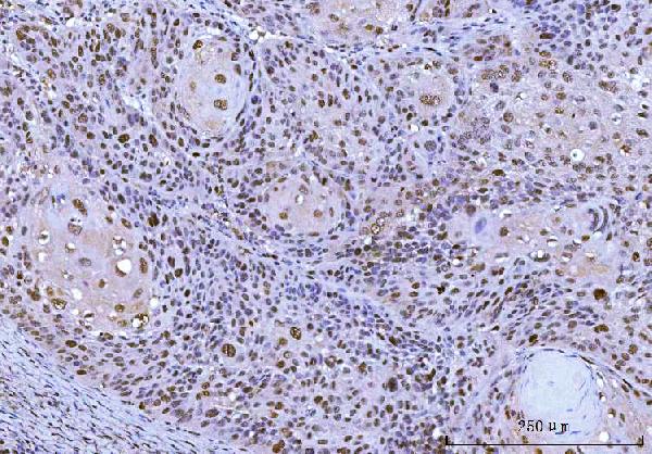
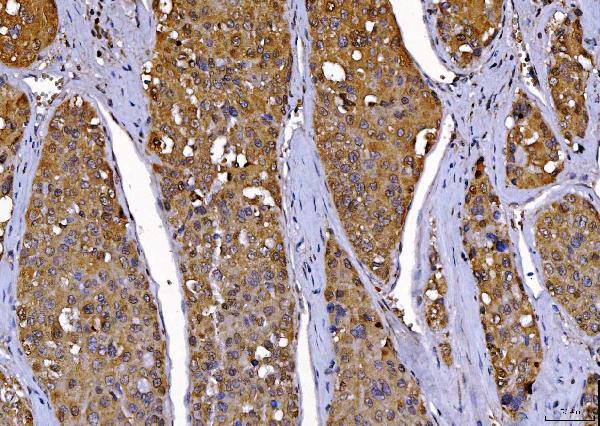
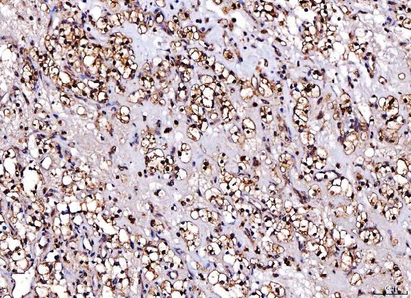
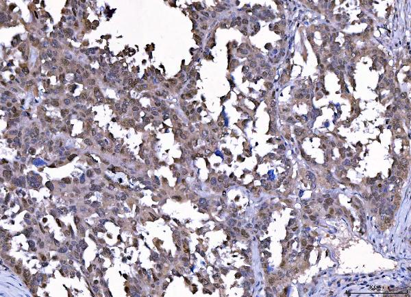
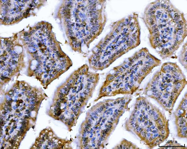
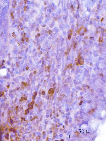
| Description | Anti-Cdk2 Antibody Picoband™ (monoclonal, 6D5B5) . Tested in IHC, WB applications. This antibody reacts with Human, Mouse, Rat. |
| Reconstitution | Adding 0.2 ml of distilled water will yield a concentration of 500 µg/ml. |
| Gene ID | 1017 |
|---|---|
| Other Names | Cyclin-dependent kinase 2, 2.7.11.22, Cell division protein kinase 2, p33 protein kinase, CDK2, CDKN2 |
| Calculated MW | 30 kDa |
| Application Details | Western blot, 0.25-0.5 µg/ml, Human, Mouse, Rat Immunohistochemistry(Paraffin-embedded Section), 2-5 µg/ml, Human, Mouse, Rat |
| Contents | Each vial contains 4 mg Trehalose, 0.9 mg NaCl and 0.2 mg Na2HPO4. |
| Clone Names | Clone: 6D5B5 |
| Immunogen | E.coli-derived human Cdk2 recombinant protein (Position: E81-L298). Human Cdk2 shares 98.6% amino acid (aa) sequence identity with rat Cdk2. |
| Purification | Immunogen affinity purified. |
| Storage | At -20°C for one year from date of receipt. After reconstitution, at 4°C for one month. It can also be aliquotted and stored frozen at -20°C for six months. Avoid repeated freezing and thawing. |
| Name | CDK2 |
|---|---|
| Synonyms | CDKN2 |
| Function | Serine/threonine-protein kinase involved in the control of the cell cycle; essential for meiosis, but dispensable for mitosis (PubMed:10499802, PubMed:10884347, PubMed:10995386, PubMed:10995387, PubMed:11051553, PubMed:11113184, PubMed:12944431, PubMed:15800615, PubMed:17495531, PubMed:19966300, PubMed:20935635, PubMed:21262353, PubMed:21596315, PubMed:28216226, PubMed:28666995). Phosphorylates CABLES1, CTNNB1, CDK2AP2, ERCC6, NBN, USP37, p53/TP53, NPM1, CDK7, RB1, BRCA2, MYC, NPAT, EZH2 (PubMed:10499802, PubMed:10995386, PubMed:10995387, PubMed:11051553, PubMed:11113184, PubMed:12944431, PubMed:15800615, PubMed:19966300, PubMed:20935635, PubMed:21262353, PubMed:21596315, PubMed:28216226). Triggers duplication of centrosomes and DNA (PubMed:11051553). Acts at the G1-S transition to promote the E2F transcriptional program and the initiation of DNA synthesis, and modulates G2 progression; controls the timing of entry into mitosis/meiosis by controlling the subsequent activation of cyclin B/CDK1 by phosphorylation, and coordinates the activation of cyclin B/CDK1 at the centrosome and in the nucleus (PubMed:18372919, PubMed:19238148, PubMed:19561645). Crucial role in orchestrating a fine balance between cellular proliferation, cell death, and DNA repair in embryonic stem cells (ESCs) (PubMed:18372919, PubMed:19238148, PubMed:19561645). Activity of CDK2 is maximal during S phase and G2; activated by interaction with cyclin E during the early stages of DNA synthesis to permit G1-S transition, and subsequently activated by cyclin A2 (cyclin A1 in germ cells) during the late stages of DNA replication to drive the transition from S phase to mitosis, the G2 phase (PubMed:18372919, PubMed:19238148, PubMed:19561645). EZH2 phosphorylation promotes H3K27me3 maintenance and epigenetic gene silencing (PubMed:20935635). Cyclin E/CDK2 prevents oxidative stress- mediated Ras-induced senescence by phosphorylating MYC (PubMed:19966300). Involved in G1-S phase DNA damage checkpoint that prevents cells with damaged DNA from initiating mitosis; regulates homologous recombination-dependent repair by phosphorylating BRCA2, this phosphorylation is low in S phase when recombination is active, but increases as cells progress towards mitosis (PubMed:15800615, PubMed:20195506, PubMed:21319273). In response to DNA damage, double- strand break repair by homologous recombination a reduction of CDK2- mediated BRCA2 phosphorylation (PubMed:15800615). Involved in regulation of telomere repair by mediating phosphorylation of NBN (PubMed:28216226). Phosphorylation of RB1 disturbs its interaction with E2F1 (PubMed:10499802). NPM1 phosphorylation by cyclin E/CDK2 promotes its dissociates from unduplicated centrosomes, thus initiating centrosome duplication (PubMed:11051553). Cyclin E/CDK2-mediated phosphorylation of NPAT at G1-S transition and until prophase stimulates the NPAT-mediated activation of histone gene transcription during S phase (PubMed:10995386, PubMed:10995387). Required for vitamin D-mediated growth inhibition by being itself inactivated (PubMed:20147522). Involved in the nitric oxide- (NO) mediated signaling in a nitrosylation/activation-dependent manner (PubMed:20079829). USP37 is activated by phosphorylation and thus triggers G1-S transition (PubMed:21596315). CTNNB1 phosphorylation regulates insulin internalization (PubMed:21262353). Phosphorylates FOXP3 and negatively regulates its transcriptional activity and protein stability (By similarity). Phosphorylates ERCC6 which is essential for its chromatin remodeling activity at DNA double-strand breaks (PubMed:29203878). Acts as a regulator of the phosphatidylinositol 3- kinase/protein kinase B signal transduction by mediating phosphorylation of the C-terminus of protein kinase B (PKB/AKT1 and PKB/AKT2), promoting its activation (PubMed:24670654). |
| Cellular Location | Cytoplasm, cytoskeleton, microtubule organizing center, centrosome. Nucleus, Cajal body. Cytoplasm. Endosome Note=Localized at the centrosomes in late G2 phase after separation of the centrosomes but before the start of prophase. Nuclear-cytoplasmic trafficking is mediated during the inhibition by 1,25-(OH)(2)D(3) |

Thousands of laboratories across the world have published research that depended on the performance of antibodies from Abcepta to advance their research. Check out links to articles that cite our products in major peer-reviewed journals, organized by research category.
info@abcepta.com, and receive a free "I Love Antibodies" mug.

Provided below are standard protocols that you may find useful for product applications.
Background
CDK2, Cyclin-Dependent Kinase2, is also known as P33. The CDK2 protein was highly homologous to p34(CDC2) kinase and more significantly homologous to Xenopus Eg1 kinase, suggesting that CDK2 is the human homolog of Eg1. The CDK2 gene is mapped to 12q13, the same region to which the CDK4 gene maps. Human cyclin A binds independently to 2 kinases, p34(cdc2) or p33. In adenovirus-transformed cells, the viral E1A oncoprotein seems to associate with p33/cyclin A but not with p34(cdc2)/cyclin A. The gene for p33 shares 65% sequence identity with p34(cdc2). P33(cdk2) plays a unique role in cell cycle regulation of vertebrate cells.
If you have used an Abcepta product and would like to share how it has performed, please click on the "Submit Review" button and provide the requested information. Our staff will examine and post your review and contact you if needed.
If you have any additional inquiries please email technical services at tech@abcepta.com.
Ordering Information
Other Products
Shipping Information
