


 Foundational characteristics of cancer include proliferation, angiogenesis, migration, evasion of apoptosis, and cellular immortality. Find key markers for these cellular processes and antibodies to detect them.
 The SUMOplot™ Analysis Program predicts and scores sumoylation sites in your protein. SUMOylation is a post-translational modification involved in various cellular processes, such as nuclear-cytosolic transport, transcriptional regulation, apoptosis, protein stability, response to stress, and progression through the cell cycle.
 The Autophagy Receptor Motif Plotter predicts and scores autophagy receptor binding sites in your protein. Identifying proteins connected to this pathway is critical to understanding the role of autophagy in physiological as well as pathological processes such as development, differentiation, neurodegenerative diseases, stress, infection, and cancer.
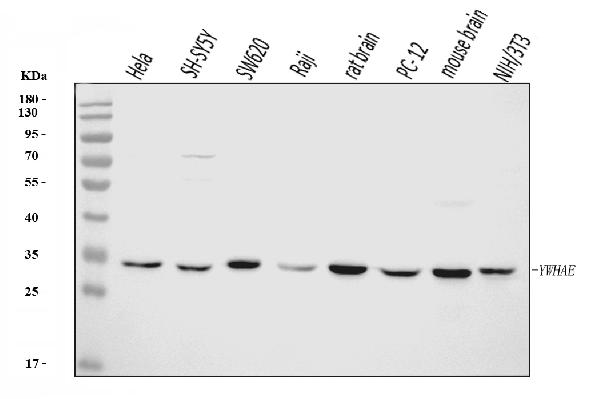
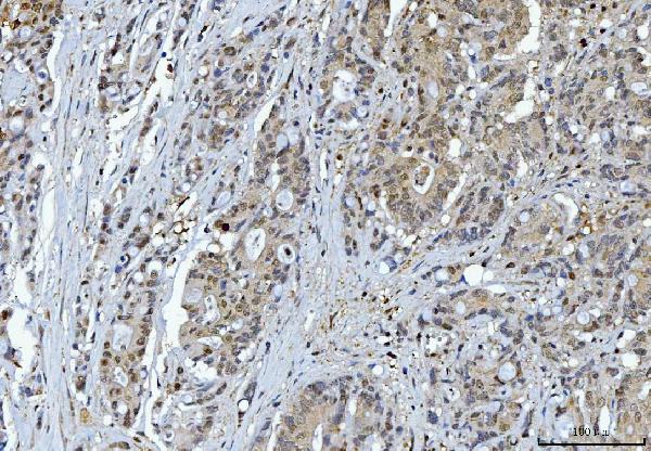
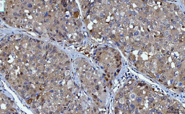
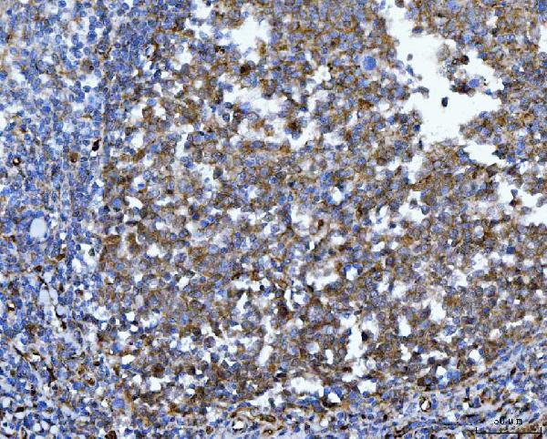
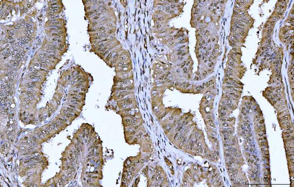
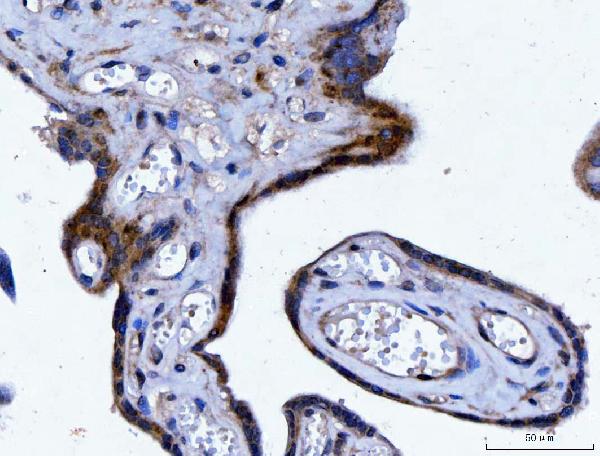
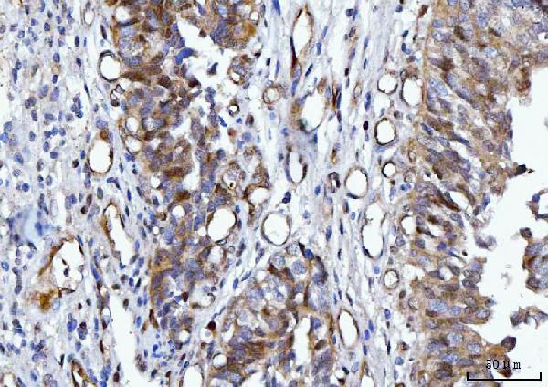
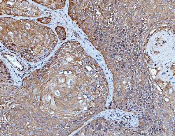
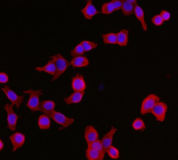
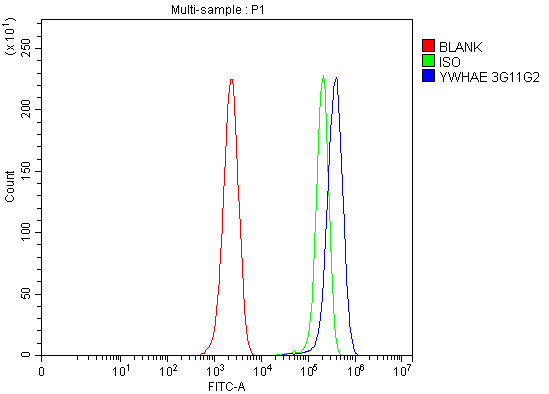
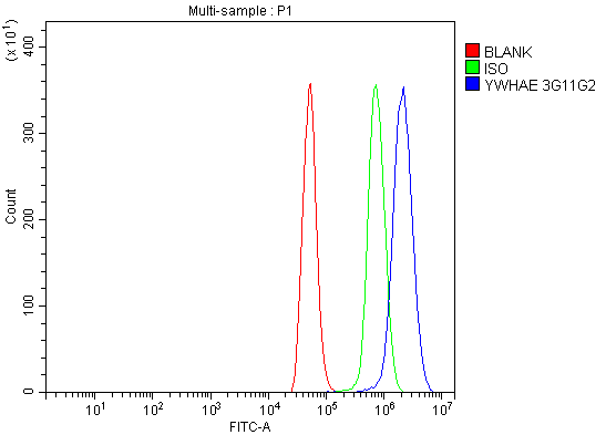
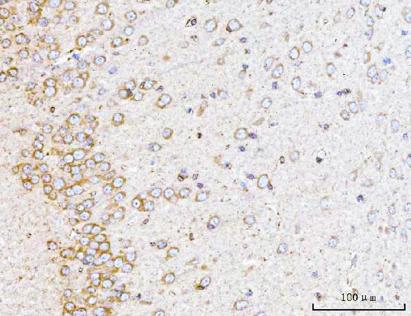
Anti-YWHAE Antibody Picoband™ (monoclonal, 3G11G2)
- SPECIFICATION
- CITATIONS
- PROTOCOLS
- BACKGROUND

Application
| WB, IHC, IF, ICC, FC |
|---|---|
| Primary Accession | P62258 |
| Host | Mouse |
| Isotype | Mouse IgG2b |
| Reactivity | Rat, Human, Mouse |
| Clonality | Monoclonal |
| Format | Lyophilized |
| Description | Anti-YWHAE Antibody Picoband™ (monoclonal, 3G11G2) . Tested in Flow Cytometry, IF, IHC, ICC, WB applications. This antibody reacts with Human, Mouse, Rat. |
| Reconstitution | Adding 0.2 ml of distilled water will yield a concentration of 500 µg/ml. |
| Gene ID | 7531 |
|---|---|
| Other Names | 14-3-3 protein epsilon, 14-3-3E, YWHAE |
| Calculated MW | 29 kDa |
| Application Details | Western blot, 0.25-0.5 µg/ml, Human, Mouse, Rat Immunohistochemistry(Paraffin-embedded Section), 2-5 µg/ml, Human, Rat Immunocytochemistry/Immunofluorescence, 5 µg/ml, Human Flow Cytometry, 1-3 µg/1x10^6 cells, Mouse, Rat |
| Contents | Each vial contains 4 mg Trehalose, 0.9 mg NaCl and 0.2 mg Na2HPO4. |
| Clone Names | Clone: 3G11G2 |
| Immunogen | E.coli-derived human YWHAE recombinant protein (Position: M1-Q255). |
| Purification | Immunogen affinity purified. |
| Storage | At -20°C for one year from date of receipt. After reconstitution, at 4°C for one month. It can also be aliquotted and stored frozen at -20°C for six months. Avoid repeated freezing and thawing. |
| Name | YWHAE |
|---|---|
| Function | Adapter protein implicated in the regulation of a large spectrum of both general and specialized signaling pathways. Binds to a large number of partners, usually by recognition of a phosphoserine or phosphothreonine motif (PubMed:35343654). Binding generally results in the modulation of the activity of the binding partner (By similarity). Positively regulates phosphorylated protein HSF1 nuclear export to the cytoplasm (PubMed:12917326). Plays a positive role in the antiviral signaling pathway upstream of TBK1 via interaction with RIGI (PubMed:37555661). Mechanistically, directs RIGI redistribution from the cytosol to mitochondrial associated membranes where it mediates MAVS-dependent innate immune signaling during viral infection (PubMed:22607805). Plays a role in proliferation inhibition and cell cycle arrest by exporting HNRNPC from the nucleus to the cytoplasm to be degraded by ubiquitination (PubMed:37599448). |
| Cellular Location | Nucleus. Cytoplasm Melanosome Note=Identified by mass spectrometry in melanosome fractions from stage I to stage IV. |

Thousands of laboratories across the world have published research that depended on the performance of antibodies from Abcepta to advance their research. Check out links to articles that cite our products in major peer-reviewed journals, organized by research category.
info@abcepta.com, and receive a free "I Love Antibodies" mug.

Provided below are standard protocols that you may find useful for product applications.
Background
14-3-3 protein epsilon is a protein that in humans is encoded by the YWHAE gene. This gene product belongs to the 14-3-3 family of proteins which mediate signal transduction by binding to phosphoserine-containing proteins. This highly conserved protein family is found in both plants and mammals, and this protein is 100% identical to the mouse ortholog. It interacts with CDC25 phosphatases, RAF1 and IRS1 proteins, suggesting its role in diverse biochemical activities related to signal transduction, such as cell division and regulation of insulin sensitivity. It has also been implicated in the pathogenesis of small cell lung cancer. Two transcript variants, one protein-coding and the other non-protein-coding, have been found for this gene.
If you have used an Abcepta product and would like to share how it has performed, please click on the "Submit Review" button and provide the requested information. Our staff will examine and post your review and contact you if needed.
If you have any additional inquiries please email technical services at tech@abcepta.com.
Ordering Information
Other Products
Shipping Information
