


 Foundational characteristics of cancer include proliferation, angiogenesis, migration, evasion of apoptosis, and cellular immortality. Find key markers for these cellular processes and antibodies to detect them.
 The SUMOplot™ Analysis Program predicts and scores sumoylation sites in your protein. SUMOylation is a post-translational modification involved in various cellular processes, such as nuclear-cytosolic transport, transcriptional regulation, apoptosis, protein stability, response to stress, and progression through the cell cycle.
 The Autophagy Receptor Motif Plotter predicts and scores autophagy receptor binding sites in your protein. Identifying proteins connected to this pathway is critical to understanding the role of autophagy in physiological as well as pathological processes such as development, differentiation, neurodegenerative diseases, stress, infection, and cancer.
Anti-FEN1 Antibody Picoband™ (monoclonal, 6F3F2)
- SPECIFICATION
- CITATIONS
- PROTOCOLS
- BACKGROUND

Application
| WB, IHC, IF, ICC, FC |
|---|---|
| Primary Accession | P39748 |
| Host | Mouse |
| Isotype | Mouse IgG1 |
| Reactivity | Rat, Human, Mouse |
| Clonality | Monoclonal |
| Format | Lyophilized |
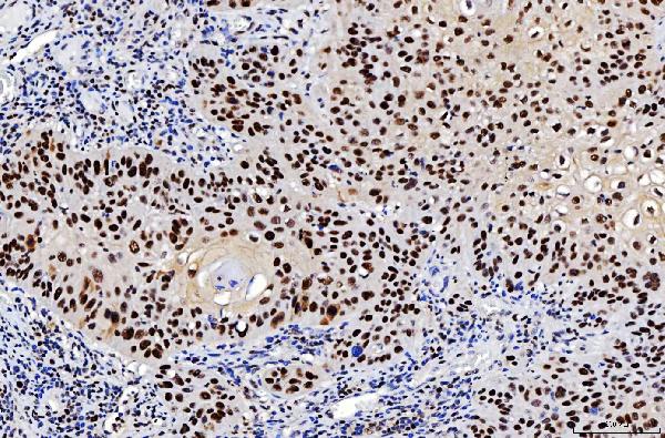
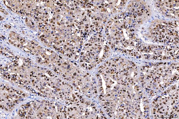
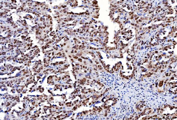
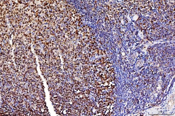
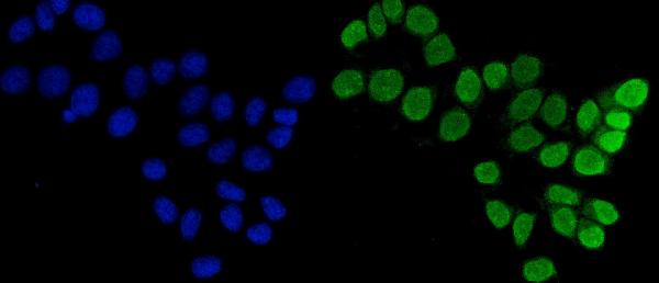
| Description | Anti-FEN1 Antibody Picoband™ (monoclonal, 6F3F2) . Tested in Flow Cytometry, IF, IHC, ICC, WB applications. This antibody reacts with Human, Mouse, Rat. |
| Reconstitution | Adding 0.2 ml of distilled water will yield a concentration of 500 µg/ml. |
| Gene ID | 2237 |
|---|---|
| Other Names | Flap endonuclease 1 {ECO:0000255|HAMAP-Rule:MF_03140}, FEN-1 {ECO:0000255|HAMAP-Rule:MF_03140}, 3.1.-.- {ECO:0000255|HAMAP-Rule:MF_03140}, DNase IV, Flap structure-specific endonuclease 1 {ECO:0000255|HAMAP-Rule:MF_03140}, Maturation factor 1, MF1, hFEN-1, FEN1 {ECO:0000255|HAMAP-Rule:MF_03140}, RAD2 |
| Calculated MW | 48 kDa |
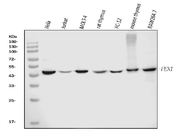
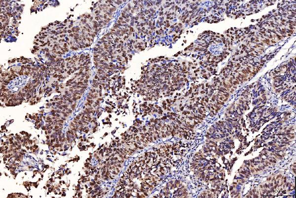
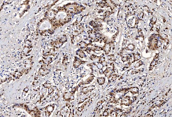
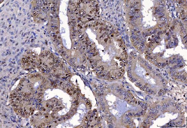
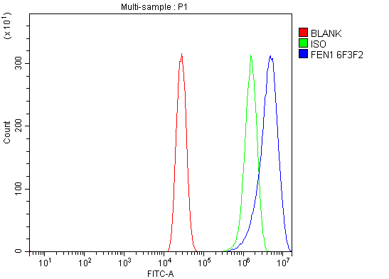
| Application Details | Western blot, 0.25-0.5 µg/ml, Human, Mouse, Rat Immunohistochemistry(Paraffin-embedded Section), 2-5 µg/ml, Human Immunocytochemistry/Immunofluorescence, 5 µg/ml, Human Flow Cytometry, 1-3 µg/1x10^6 cells, Human |
| Contents | Each vial contains 4 mg Trehalose, 0.9 mg NaCl and 0.2 mg Na2HPO4. |
| Clone Names | Clone: 6F3F2 |
| Immunogen | E.coli-derived human FEN1 recombinant protein (Position: Q4-E300). |
| Purification | Immunogen affinity purified. |
| Storage | At -20°C for one year from date of receipt. After reconstitution, at 4°C for one month. It can also be aliquotted and stored frozen at -20°C for six months. Avoid repeated freezing and thawing. |
| Name | FEN1 {ECO:0000255|HAMAP-Rule:MF_03140} |
|---|---|
| Synonyms | RAD2 |
| Function | Structure-specific nuclease with 5'-flap endonuclease and 5'- 3' exonuclease activities involved in DNA replication and repair. During DNA replication, cleaves the 5'-overhanging flap structure that is generated by displacement synthesis when DNA polymerase encounters the 5'-end of a downstream Okazaki fragment. It enters the flap from the 5'-end and then tracks to cleave the flap base, leaving a nick for ligation. Also involved in the long patch base excision repair (LP-BER) pathway, by cleaving within the apurinic/apyrimidinic (AP) site- terminated flap. Acts as a genome stabilization factor that prevents flaps from equilibrating into structures that lead to duplications and deletions. Also possesses 5'-3' exonuclease activity on nicked or gapped double-stranded DNA, and exhibits RNase H activity. Also involved in replication and repair of rDNA and in repairing mitochondrial DNA. |
| Cellular Location | [Isoform 1]: Nucleus, nucleolus. Nucleus, nucleoplasm. Note=Resides mostly in the nucleoli and relocalizes to the nucleoplasm upon DNA damage |

Thousands of laboratories across the world have published research that depended on the performance of antibodies from Abcepta to advance their research. Check out links to articles that cite our products in major peer-reviewed journals, organized by research category.
info@abcepta.com, and receive a free "I Love Antibodies" mug.

Provided below are standard protocols that you may find useful for product applications.
Background
Flap endonuclease 1 is an enzyme that in humans is encoded by the FEN1 gene. It is mapped to 11q12.2. The protein encoded by this gene removes 5' overhanging flaps in DNA repair and processes the 5' ends of Okazaki fragments in lagging strand DNA synthesis. Direct physical interaction between this protein and AP endonuclease 1 during long-patch base excision repair provides coordinated loading of the proteins onto the substrate, thus passing the substrate from one enzyme to another. The protein is a member of the XPG/RAD2 endonuclease family and is one of ten proteins essential for cell-free DNA replication. DNA secondary structure can inhibit flap processing at certain trinucleotide repeats in a length-dependent manner by concealing the 5' end of the flap that is necessary for both binding and cleavage by the protein encoded by this gene. Therefore, secondary structure can deter the protective function of this protein, leading to site-specific trinucleotide expansions.
If you have used an Abcepta product and would like to share how it has performed, please click on the "Submit Review" button and provide the requested information. Our staff will examine and post your review and contact you if needed.
If you have any additional inquiries please email technical services at tech@abcepta.com.
Ordering Information
Other Products
Shipping Information
