


 Foundational characteristics of cancer include proliferation, angiogenesis, migration, evasion of apoptosis, and cellular immortality. Find key markers for these cellular processes and antibodies to detect them.
 The SUMOplot™ Analysis Program predicts and scores sumoylation sites in your protein. SUMOylation is a post-translational modification involved in various cellular processes, such as nuclear-cytosolic transport, transcriptional regulation, apoptosis, protein stability, response to stress, and progression through the cell cycle.
 The Autophagy Receptor Motif Plotter predicts and scores autophagy receptor binding sites in your protein. Identifying proteins connected to this pathway is critical to understanding the role of autophagy in physiological as well as pathological processes such as development, differentiation, neurodegenerative diseases, stress, infection, and cancer.
Anti-SHP1/PTPN6 Antibody Picoband™ (monoclonal, 8H11B10)
- SPECIFICATION
- CITATIONS
- PROTOCOLS
- BACKGROUND

Application
| WB, IHC, IF, ICC, FC |
|---|---|
| Primary Accession | P29350 |
| Host | Mouse |
| Isotype | Mouse IgG2b |
| Reactivity | Rat, Human, Mouse |
| Clonality | Monoclonal |
| Format | Lyophilized |
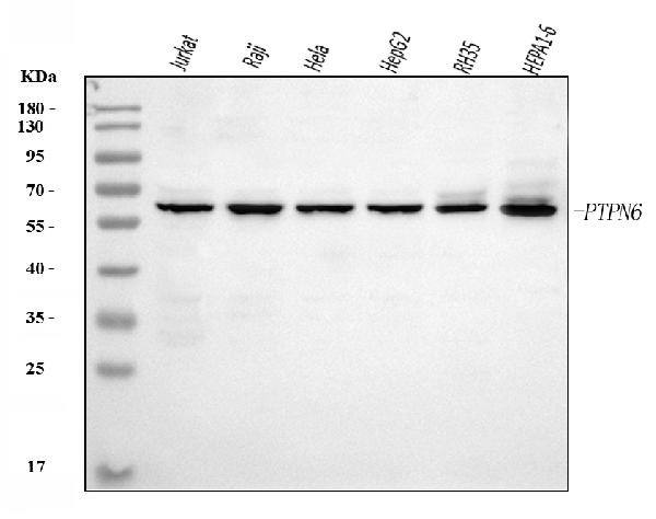
| Description | Anti-SHP1/PTPN6 Antibody Picoband™ (monoclonal, 8H11B10) . Tested in Flow Cytometry, IF, IHC, ICC, WB applications. This antibody reacts with Human, Mouse, Rat. |
| Reconstitution | Adding 0.2 ml of distilled water will yield a concentration of 500 µg/ml. |
| Gene ID | 5777 |
|---|---|
| Other Names | Tyrosine-protein phosphatase non-receptor type 6, 3.1.3.48, Hematopoietic cell protein-tyrosine phosphatase, Protein-tyrosine phosphatase 1C, PTP-1C, Protein-tyrosine phosphatase SHP-1, SH-PTP1, PTPN6, HCP, PTP1C |
| Calculated MW | 68 kDa |
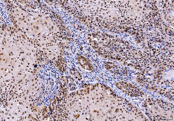
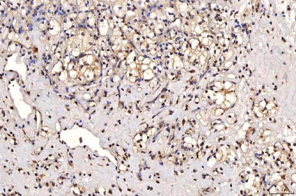
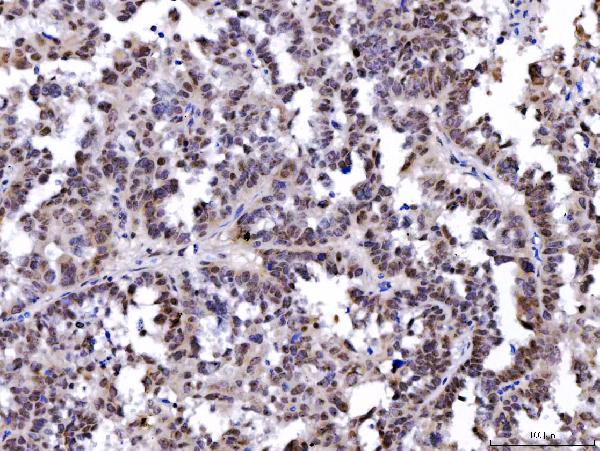
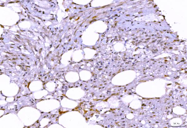
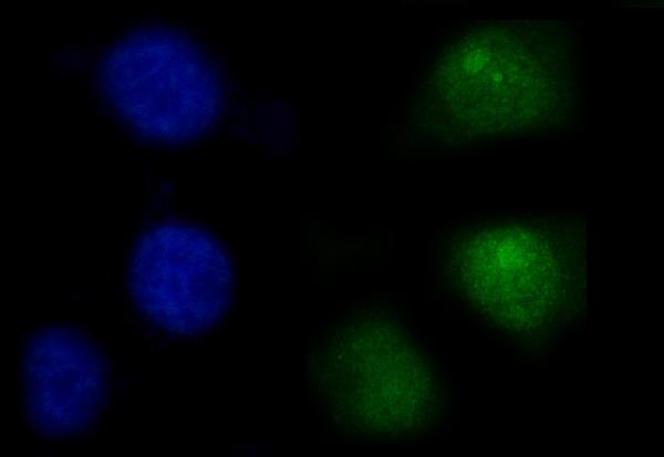
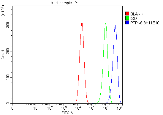
| Application Details | Western blot, 0.25-0.5 µg/ml, Human, Mouse, Rat Immunohistochemistry(Paraffin-embedded Section), 2-5 µg/ml, Human Immunocytochemistry/Immunofluorescence, 5 µg/ml, Human Flow Cytometry, 1-3 µg/1x10^6 cells, Human |
| Contents | Each vial contains 4 mg Trehalose, 0.9 mg NaCl and 0.2 mg Na2HPO4. |
| Clone Names | Clone: 8H11B10 |
| Immunogen | E.coli-derived human SHP1/PTPN6 recombinant protein (Position: E67-K572). |
| Purification | Immunogen affinity purified. |
| Storage | At -20°C for one year from date of receipt. After reconstitution, at 4°C for one month. It can also be aliquotted and stored frozen at -20°C for six months. Avoid repeated freezing and thawing. |
| Name | PTPN6 |
|---|---|
| Synonyms | HCP, PTP1C |
| Function | Tyrosine phosphatase enzyme that plays important roles in controlling immune signaling pathways and fundamental physiological processes such as hematopoiesis (PubMed:14739280, PubMed:29925997). Dephosphorylates and negatively regulate several receptor tyrosine kinases (RTKs) such as EGFR, PDGFR and FGFR, thereby modulating their signaling activities (PubMed:21258366, PubMed:9733788). When recruited to immunoreceptor tyrosine-based inhibitory motif (ITIM)-containing receptors such as immunoglobulin-like transcript 2/LILRB1, programmed cell death protein 1/PDCD1, CD3D, CD22, CLEC12A and other receptors involved in immune regulation, initiates their dephosphorylation and subsequently inhibits downstream signaling events (PubMed:11907092, PubMed:14739280, PubMed:37932456, PubMed:38166031). Modulates the signaling of several cytokine receptors including IL-4 receptor (PubMed:9065461). Additionally, targets multiple cytoplasmic signaling molecules including STING1, LCK or STAT1 among others involved in diverse cellular processes including modulation of T-cell activation or cGAS-STING signaling (PubMed:34811497, PubMed:38532423). Within the nucleus, negatively regulates the activity of some transcription factors such as NFAT5 via direct dephosphorylation. Acts also as a key transcriptional regulator of hepatic gluconeogenesis by controlling recruitment of RNA polymerase II to the PCK1 promoter together with STAT5A (PubMed:37595871). |
| Cellular Location | Cytoplasm. Nucleus Note=In neurons, translocates into the nucleus after treatment with angiotensin II (By similarity). Shuttles between the cytoplasm and nucleus via its association with PDPK1. |
| Tissue Location | Isoform 1 is expressed in hematopoietic cells. Isoform 2 is expressed in non-hematopoietic cells |

Thousands of laboratories across the world have published research that depended on the performance of antibodies from Abcepta to advance their research. Check out links to articles that cite our products in major peer-reviewed journals, organized by research category.
info@abcepta.com, and receive a free "I Love Antibodies" mug.

Provided below are standard protocols that you may find useful for product applications.
Background
Tyrosine-protein phosphatase non-receptor type 6, also known as Src homology region 2 domain-containing phosphatase-1 (SHP-1), is an enzyme that in humans is encoded by the PTPN6 gene. The protein encoded by this gene is a member of the protein tyrosine phosphatase (PTP) family. PTPs are known to be signaling molecules that regulate a variety of cellular processes including cell growth, differentiation, mitotic cycle, and oncogenic transformation. N-terminal part of this PTP contains two tandem Src homolog (SH2) domains, which act as protein phospho-tyrosine binding domains, and mediate the interaction of this PTP with its substrates. This PTP is expressed primarily in hematopoietic cells, and functions as an important regulator of multiple signaling pathways in hematopoietic cells. This PTP has been shown to interact with, and dephosphorylate a wide spectrum of phospho-proteins involved in hematopoietic cell signaling. Multiple alternatively spliced variants of this gene, which encode distinct isoforms, have been reported.
If you have used an Abcepta product and would like to share how it has performed, please click on the "Submit Review" button and provide the requested information. Our staff will examine and post your review and contact you if needed.
If you have any additional inquiries please email technical services at tech@abcepta.com.
Ordering Information
Other Products
Shipping Information
