


 Foundational characteristics of cancer include proliferation, angiogenesis, migration, evasion of apoptosis, and cellular immortality. Find key markers for these cellular processes and antibodies to detect them.
 The SUMOplot™ Analysis Program predicts and scores sumoylation sites in your protein. SUMOylation is a post-translational modification involved in various cellular processes, such as nuclear-cytosolic transport, transcriptional regulation, apoptosis, protein stability, response to stress, and progression through the cell cycle.
 The Autophagy Receptor Motif Plotter predicts and scores autophagy receptor binding sites in your protein. Identifying proteins connected to this pathway is critical to understanding the role of autophagy in physiological as well as pathological processes such as development, differentiation, neurodegenerative diseases, stress, infection, and cancer.
> home > Products > Primary Antibodies > Signal Transduction > Anti-PPP6C Rabbit Monoclonal Antibody
Anti-PPP6C Rabbit Monoclonal Antibody
- SPECIFICATION
- CITATIONS
- PROTOCOLS
- BACKGROUND

Application
| WB, IF, ICC, FC |
|---|---|
| Primary Accession | O00743 |
| Host | Rabbit |
| Isotype | IgG |
| Reactivity | Human |
| Clonality | Monoclonal |
| Format | Liquid |
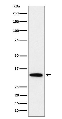
| Description | Anti-PPP6C Rabbit Monoclonal Antibody . Tested in WB, ICC/IF, Flow Cytometry applications. This antibody reacts with Human. |
| Gene ID | 5537 |
|---|---|
| Other Names | Serine/threonine-protein phosphatase 6 catalytic subunit, PP6C, 3.1.3.16, Serine/threonine-protein phosphatase 6 catalytic subunit, N-terminally processed, PPP6C {ECO:0000303|PubMed:29053956, ECO:0000312|HGNC:HGNC:9323} |
| Calculated MW | 32 kDa |
| Application Details | WB 1:500-1:2000 ICC/IF 1:50-1:200 FC 1:50 |
| Contents | Rabbit IgG in phosphate buffered saline, pH 7.4, 150mM NaCl, 0.02% sodium azide and 50% glycerol, 0.4-0.5mg/ml BSA. |
| Clone Names | Clone: 27P53 |
| Immunogen | A synthesized peptide derived from human PPP6C |
| Purification | Affinity-chromatography |
| Storage | Store at -20°C for one year. For short term storage and frequent use, store at 4°C for up to one month. Avoid repeated freeze-thaw cycles. |
| Name | PPP6C {ECO:0000303|PubMed:29053956, ECO:0000312|HGNC:HGNC:9323} |
|---|---|
| Function | Catalytic subunit of protein phosphatase 6 (PP6) (PubMed:17079228, PubMed:29053956, PubMed:32474700). PP6 is a component of a signaling pathway regulating cell cycle progression in response to IL2 receptor stimulation (PubMed:10227379). N-terminal domain restricts G1 to S phase progression in cancer cells, in part through control of cyclin D1 (PubMed:17568194). During mitosis, regulates spindle positioning (PubMed:27335426). Down-regulates MAP3K7 kinase activation of the IL1 signaling pathway by dephosphorylation of MAP3K7 (PubMed:17079228). Participates also in the innate immune defense against viruses by desphosphorylating RIGI, an essential step that triggers RIGI-mediated signaling activation (PubMed:29053956). Also regulates innate immunity by acting as a negative regulator of the cGAS-STING pathway: mediates dephosphorylation and inactivation of CGAS and STING1 (PubMed:32474700, PubMed:32753499). CGAS dephosphorylation at 'Ser-435' impairs its ability to bind GTP, thereby inactivating it (PubMed:32474700). |
| Cellular Location | Mitochondrion. Cytoplasm |
| Tissue Location | Ubiquitously expressed in all tissues tested with highest expression levels in testis, heart, kidney, brain, stomach, liver and skeletal muscle and lowest in placenta, lung colon and spleen. |
Research Areas
Citations (0)

Thousands of laboratories across the world have published research that depended on the performance of antibodies from Abcepta to advance their research. Check out links to articles that cite our products in major peer-reviewed journals, organized by research category.
Submit your citation using an Abcepta antibody to
info@abcepta.com, and receive a free "I Love Antibodies" mug.
info@abcepta.com, and receive a free "I Love Antibodies" mug.

Application Protocols
Provided below are standard protocols that you may find useful for product applications.
Abcepta welcomes feedback from its customers.
If you have used an Abcepta product and would like to share how it has performed, please click on the "Submit Review" button and provide the requested information. Our staff will examine and post your review and contact you if needed.
If you have any additional inquiries please email technical services at tech@abcepta.com.
$ 370.00
Cat# ABO16174
Ordering Information
United States
AlbaniaAustraliaAustriaBelgiumBosnia & HerzegovinaBrazilBulgariaCanadaCentral AmericaChinaCroatiaCyprusCzech RepublicDenmarkEstoniaFinlandFranceGermanyGreeceHong KongHungaryIcelandIndiaIndonesiaIrelandIsraelItalyJapanLatviaLithuaniaLuxembourgMacedoniaMalaysiaMaltaNetherlandsNew ZealandNorwayPakistanPolandPortugalRomaniaSerbiaSingaporeSlovakiaSloveniaSouth AfricaSouth KoreaSpainSwedenSwitzerlandTaiwanTurkeyUnited KingdomUnited StatesVietnamWorldwideOthersMexico
USA Headquarters
(888) 735-7227 / (858) 622-0099 or (858) 875-1900
Other Products
Shipping Information
Domestic orders (in stock items)
Shipped out the same day. Orders placed after 1 PM (PST) will ship out the next business day.
International orders
Contact your local distributors
