


 Foundational characteristics of cancer include proliferation, angiogenesis, migration, evasion of apoptosis, and cellular immortality. Find key markers for these cellular processes and antibodies to detect them.
 The SUMOplot™ Analysis Program predicts and scores sumoylation sites in your protein. SUMOylation is a post-translational modification involved in various cellular processes, such as nuclear-cytosolic transport, transcriptional regulation, apoptosis, protein stability, response to stress, and progression through the cell cycle.
 The Autophagy Receptor Motif Plotter predicts and scores autophagy receptor binding sites in your protein. Identifying proteins connected to this pathway is critical to understanding the role of autophagy in physiological as well as pathological processes such as development, differentiation, neurodegenerative diseases, stress, infection, and cancer.
Anti-ACVR1B Rabbit Monoclonal Antibody
- SPECIFICATION
- CITATIONS
- PROTOCOLS
- BACKGROUND

Application
| WB, IHC, IF, ICC |
|---|---|
| Primary Accession | P36896 |
| Host | Rabbit |
| Isotype | IgG |
| Reactivity | Rat, Human, Mouse |
| Clonality | Monoclonal |
| Format | Liquid |
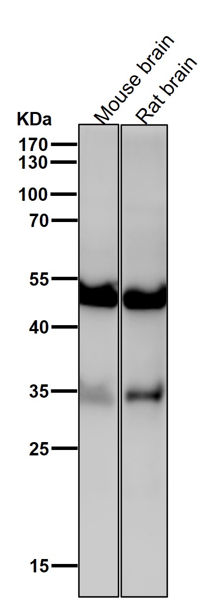
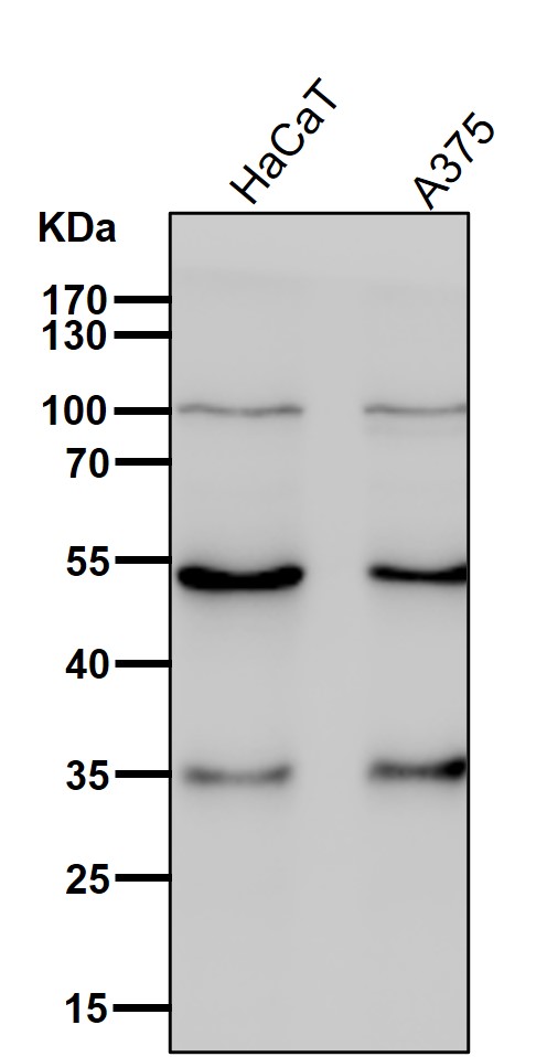
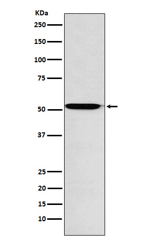
| Description | Anti-ACVR1B Rabbit Monoclonal Antibody . Tested in WB, IHC, ICC/IF applications. This antibody reacts with Human, Mouse, Rat. |
| Gene ID | 91 |
|---|---|
| Other Names | Activin receptor type-1B, 2.7.11.30, Activin receptor type IB, ACTR-IB, Activin receptor-like kinase 4, ALK-4, Serine/threonine-protein kinase receptor R2, SKR2, ACVR1B, ACVRLK4, ALK4 |
| Calculated MW | 52 kDa |
| Application Details | WB 1:500-1:2000 IHC 1:100-1:500 ICC/IF 1:50-1:200 |
| Contents | Rabbit IgG in phosphate buffered saline, pH 7.4, 150mM NaCl, 0.02% sodium azide and 50% glycerol, 0.4-0.5mg/ml BSA. |
| Clone Names | Clone: 23A40 |
| Immunogen | A synthesized peptide derived from human ACVR1B |
| Purification | Affinity-chromatography |
| Storage | Store at -20°C for one year. For short term storage and frequent use, store at 4°C for up to one month. Avoid repeated freeze-thaw cycles. |
| Name | ACVR1B |
|---|---|
| Synonyms | ACVRLK4, ALK4 |
| Function | Transmembrane serine/threonine kinase activin type-1 receptor forming an activin receptor complex with activin receptor type-2 (ACVR2A or ACVR2B). Transduces the activin signal from the cell surface to the cytoplasm and is thus regulating a many physiological and pathological processes including neuronal differentiation and neuronal survival, hair follicle development and cycling, FSH production by the pituitary gland, wound healing, extracellular matrix production, immunosuppression and carcinogenesis. Activin is also thought to have a paracrine or autocrine role in follicular development in the ovary. Within the receptor complex, type-2 receptors (ACVR2A and/or ACVR2B) act as a primary activin receptors whereas the type-1 receptors like ACVR1B act as downstream transducers of activin signals. Activin binds to type-2 receptor at the plasma membrane and activates its serine- threonine kinase. The activated receptor type-2 then phosphorylates and activates the type-1 receptor such as ACVR1B. Once activated, the type- 1 receptor binds and phosphorylates the SMAD proteins SMAD2 and SMAD3, on serine residues of the C-terminal tail. Soon after their association with the activin receptor and subsequent phosphorylation, SMAD2 and SMAD3 are released into the cytoplasm where they interact with the common partner SMAD4. This SMAD complex translocates into the nucleus where it mediates activin-induced transcription. Inhibitory SMAD7, which is recruited to ACVR1B through FKBP1A, can prevent the association of SMAD2 and SMAD3 with the activin receptor complex, thereby blocking the activin signal. Activin signal transduction is also antagonized by the binding to the receptor of inhibin-B via the IGSF1 inhibin coreceptor. ACVR1B also phosphorylates TDP2. |
| Cellular Location | Cell membrane; Single-pass type I membrane protein |
| Tissue Location | Expressed in many tissues, most strongly in kidney, pancreas, brain, lung, and liver |
Research Areas
Citations (0)

Thousands of laboratories across the world have published research that depended on the performance of antibodies from Abcepta to advance their research. Check out links to articles that cite our products in major peer-reviewed journals, organized by research category.
Submit your citation using an Abcepta antibody to
info@abcepta.com, and receive a free "I Love Antibodies" mug.
info@abcepta.com, and receive a free "I Love Antibodies" mug.

Application Protocols
Provided below are standard protocols that you may find useful for product applications.
Abcepta welcomes feedback from its customers.
If you have used an Abcepta product and would like to share how it has performed, please click on the "Submit Review" button and provide the requested information. Our staff will examine and post your review and contact you if needed.
If you have any additional inquiries please email technical services at tech@abcepta.com.
$ 370.00
Cat# ABO15764
Ordering Information
United States
AlbaniaAustraliaAustriaBelgiumBosnia & HerzegovinaBrazilBulgariaCanadaCentral AmericaChinaCroatiaCyprusCzech RepublicDenmarkEstoniaFinlandFranceGermanyGreeceHong KongHungaryIcelandIndiaIndonesiaIrelandIsraelItalyJapanLatviaLithuaniaLuxembourgMacedoniaMalaysiaMaltaMexicoNetherlandsNew ZealandNorwayPakistanPolandPortugalRomaniaSerbiaSingaporeSlovakiaSloveniaSouth AfricaSouth KoreaSpainSwedenSwitzerlandTaiwanTurkeyUnited KingdomUnited StatesVietnamWorldwideOthers
USA Headquarters
(888) 735-7227 / (858) 622-0099 or (858) 875-1900
Other Products
Shipping Information
Domestic orders (in stock items)
Shipped out the same day. Orders placed after 1 PM (PST) will ship out the next business day.
International orders
Contact your local distributors
