Anti-PAR2 Rabbit Monoclonal Antibody
- SPECIFICATION
- CITATIONS
- PROTOCOLS
- BACKGROUND

Application
| WB, IHC, IF, ICC, FC |
|---|---|
| Primary Accession | P55085 |
| Host | Rabbit |
| Isotype | IgG |
| Reactivity | Rat, Human, Mouse |
| Clonality | Monoclonal |
| Format | Liquid |
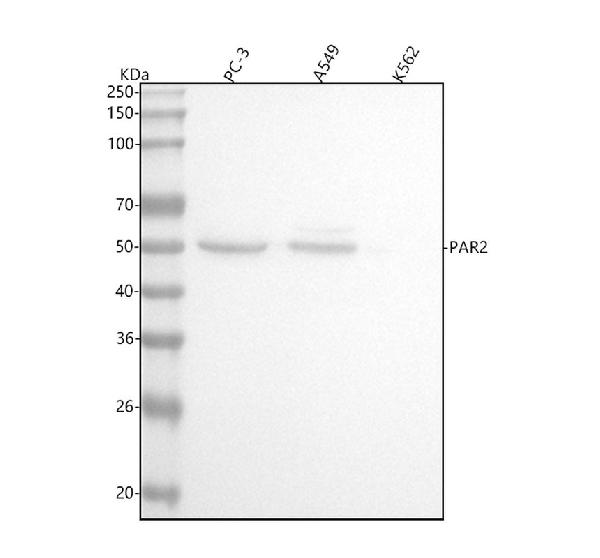
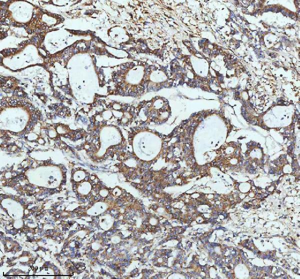
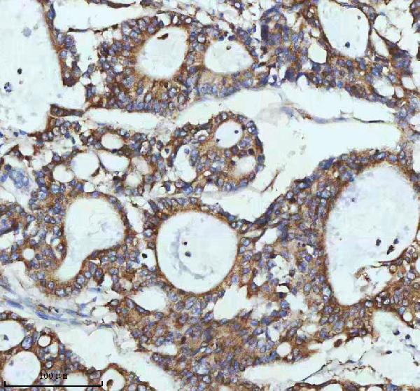
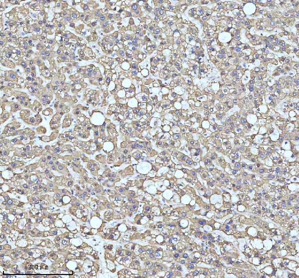
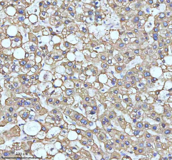
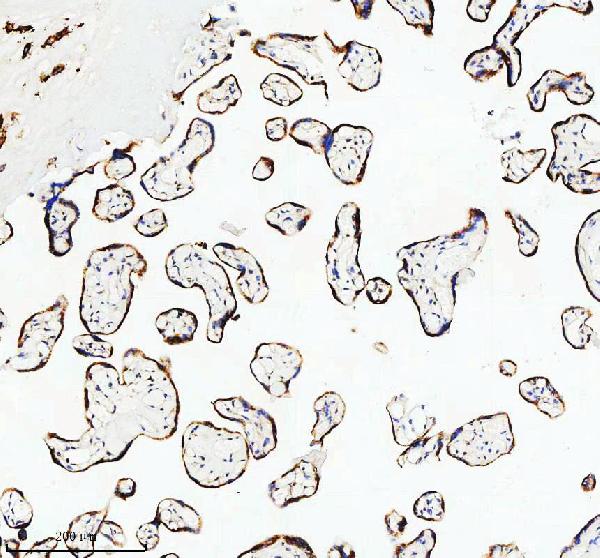
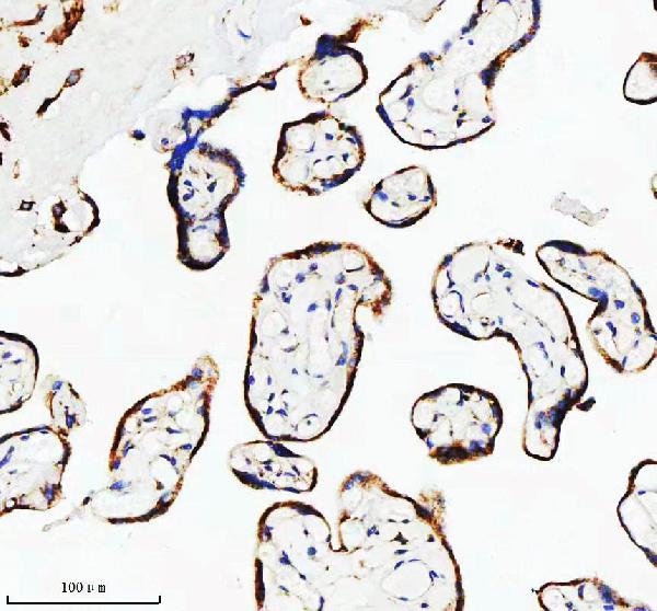
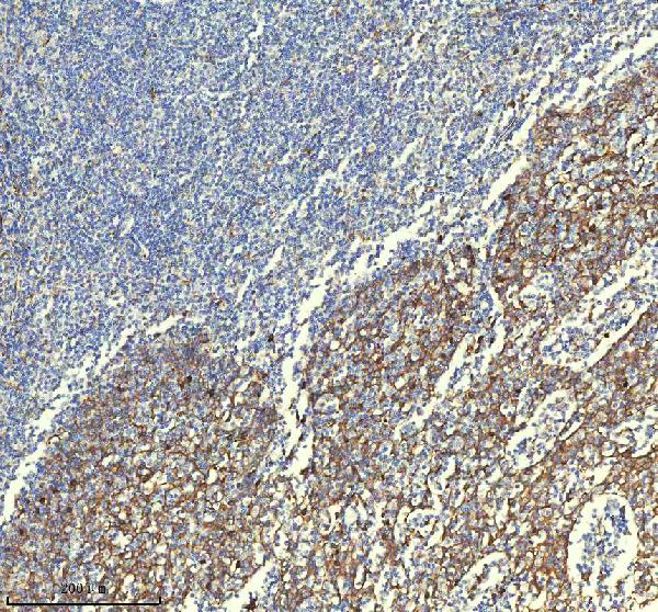
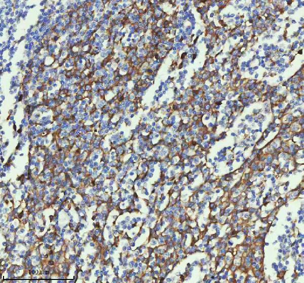
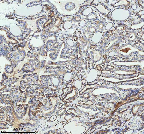
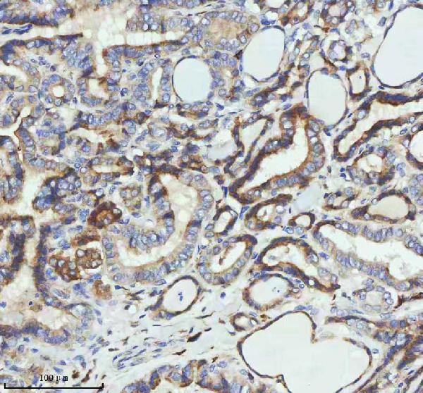
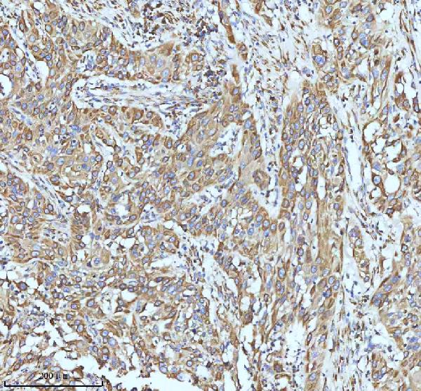
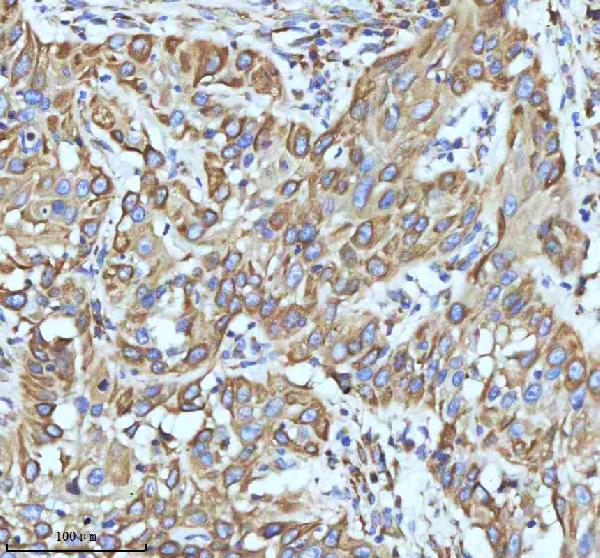
| Description | Anti-PAR2 Rabbit Monoclonal Antibody . Tested in WB, IHC, ICC/IF, Flow Cytometry applications. This antibody reacts with Human, Mouse, Rat. |
| Gene ID | 2150 |
|---|---|
| Other Names | Proteinase-activated receptor 2, PAR-2, Coagulation factor II receptor-like 1, G-protein coupled receptor 11, Thrombin receptor-like 1, Proteinase-activated receptor 2, alternate cleaved 1, Proteinase-activated receptor 2, alternate cleaved 2, F2RL1, GPR11, PAR2 |
| Calculated MW | 55 kDa |
| Application Details | WB 1:500-1:2000 IHC 1:50-1:200 ICC/IF 1:50-1:200 FC 1:50 |
| Contents | Rabbit IgG in phosphate buffered saline, pH 7.4, 150mM NaCl, 0.02% sodium azide and 50% glycerol, 0.4-0.5mg/ml BSA. |
| Clone Names | Clone: 22F95 |
| Immunogen | A synthesized peptide derived from human PAR2 |
| Purification | Affinity-chromatography |
| Storage | Store at -20°C for one year. For short term storage and frequent use, store at 4°C for up to one month. Avoid repeated freeze-thaw cycles. |
| Name | F2RL1 |
|---|---|
| Synonyms | GPR11, PAR2 |
| Function | Receptor for trypsin and trypsin-like enzymes coupled to G proteins (PubMed:28445455). Its function is mediated through the activation of several signaling pathways including phospholipase C (PLC), intracellular calcium, mitogen-activated protein kinase (MAPK), I-kappaB kinase/NF-kappaB and Rho (PubMed:28445455). Can also be transactivated by cleaved F2R/PAR1. Involved in modulation of inflammatory responses and regulation of innate and adaptive immunity, and acts as a sensor for proteolytic enzymes generated during infection. Generally is promoting inflammation. Can signal synergistically with TLR4 and probably TLR2 in inflammatory responses and modulates TLR3 signaling. Has a protective role in establishing the endothelial barrier; the activity involves coagulation factor X. Regulates endothelial cell barrier integrity during neutrophil extravasation, probably following proteolytic cleavage by PRTN3 (PubMed:23202369). Proposed to have a bronchoprotective role in airway epithelium, but also shown to compromise the airway epithelial barrier by interrupting E-cadherin adhesion (PubMed:10086357). Involved in the regulation of vascular tone; activation results in hypotension presumably mediated by vasodilation. Associates with a subset of G proteins alpha subunits such as GNAQ, GNA11, GNA14, GNA12 and GNA13, but probably not with G(o)-alpha, G(i) subunit alpha-1 and G(i) subunit alpha-2. However, according to PubMed:21627585 can signal through G(i) subunit alpha. Believed to be a class B receptor which internalizes as a complex with arrestin and traffic with it to endosomal vesicles, presumably as desensitized receptor, for extended periods of time. Mediates inhibition of TNF-alpha stimulated JNK phosphorylation via coupling to GNAQ and GNA11; the function involves dissociation of RIPK1 and TRADD from TNFR1. Mediates phosphorylation of nuclear factor NF- kappa-B RELA subunit at 'Ser-536'; the function involves IKBKB and is predominantly independent of G proteins. Involved in cellular migration. Involved in cytoskeletal rearrangement and chemotaxis through beta-arrestin-promoted scaffolds; the function is independent of GNAQ and GNA11 and involves promotion of cofilin dephosphorylation and actin filament severing. Induces redistribution of COPS5 from the plasma membrane to the cytosol and activation of the JNK cascade is mediated by COPS5. Involved in the recruitment of leukocytes to the sites of inflammation and is the major PAR receptor capable of modulating eosinophil function such as pro-inflammatory cytokine secretion, superoxide production and degranulation. During inflammation promotes dendritic cell maturation, trafficking to the lymph nodes and subsequent T-cell activation. Involved in antimicrobial response of innate immune cells; activation enhances phagocytosis of Gram-positive and killing of Gram-negative bacteria. Acts synergistically with interferon-gamma in enhancing antiviral responses. Implicated in a number of acute and chronic inflammatory diseases such as of the joints, lungs, brain, gastrointestinal tract, periodontium, skin, and vascular systems, and in autoimmune disorders. Probably mediates activation of pro-inflammatory and pro-fibrotic responses in fibroblasts, triggered by coagulation factor Xa (F10) (By similarity). Mediates activation of barrier protective signaling responses in endothelial cells, triggered by coagulation factor Xa (F10) (PubMed:22409427). |
| Cellular Location | Cell membrane; Multi-pass membrane protein. |
| Tissue Location | Widely expressed in tissues with especially high levels in pancreas, liver, kidney, small intestine, and colon (PubMed:7556175, PubMed:8615752). Moderate expression is detected in many organs, but none in brain or skeletal muscle (PubMed:7556175, PubMed:8615752). Expressed in endothelial cells (PubMed:23202369) |

Thousands of laboratories across the world have published research that depended on the performance of antibodies from Abcepta to advance their research. Check out links to articles that cite our products in major peer-reviewed journals, organized by research category.
info@abcepta.com, and receive a free "I Love Antibodies" mug.
Provided below are standard protocols that you may find useful for product applications.
If you have used an Abcepta product and would like to share how it has performed, please click on the "Submit Review" button and provide the requested information. Our staff will examine and post your review and contact you if needed.
If you have any additional inquiries please email technical services at tech@abcepta.com.


 Foundational characteristics of cancer include proliferation, angiogenesis, migration, evasion of apoptosis, and cellular immortality. Find key markers for these cellular processes and antibodies to detect them.
 The SUMOplot™ Analysis Program predicts and scores sumoylation sites in your protein. SUMOylation is a post-translational modification involved in various cellular processes, such as nuclear-cytosolic transport, transcriptional regulation, apoptosis, protein stability, response to stress, and progression through the cell cycle.
 The Autophagy Receptor Motif Plotter predicts and scores autophagy receptor binding sites in your protein. Identifying proteins connected to this pathway is critical to understanding the role of autophagy in physiological as well as pathological processes such as development, differentiation, neurodegenerative diseases, stress, infection, and cancer.
