


 Foundational characteristics of cancer include proliferation, angiogenesis, migration, evasion of apoptosis, and cellular immortality. Find key markers for these cellular processes and antibodies to detect them.
 The SUMOplot™ Analysis Program predicts and scores sumoylation sites in your protein. SUMOylation is a post-translational modification involved in various cellular processes, such as nuclear-cytosolic transport, transcriptional regulation, apoptosis, protein stability, response to stress, and progression through the cell cycle.
 The Autophagy Receptor Motif Plotter predicts and scores autophagy receptor binding sites in your protein. Identifying proteins connected to this pathway is critical to understanding the role of autophagy in physiological as well as pathological processes such as development, differentiation, neurodegenerative diseases, stress, infection, and cancer.
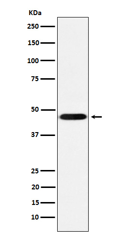
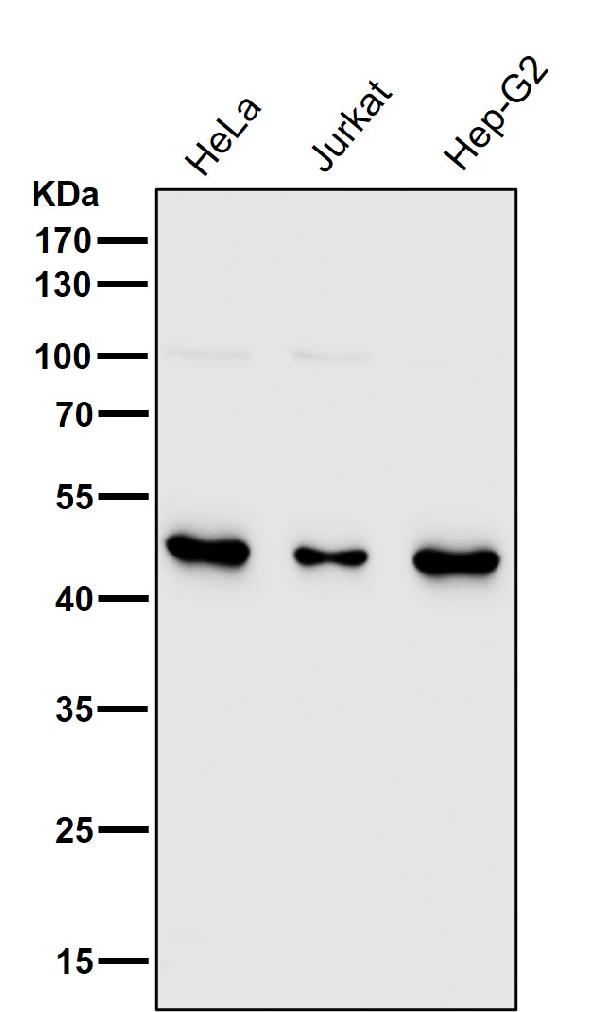
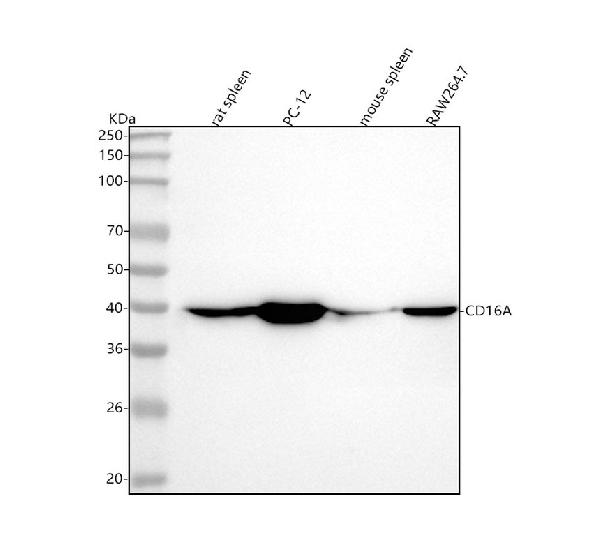
Anti-CD16 Rabbit Monoclonal Antibody
- SPECIFICATION
- CITATIONS
- PROTOCOLS
- BACKGROUND

Application
| WB, IF, ICC, FC |
|---|---|
| Primary Accession | P08637 |
| Host | Rabbit |
| Isotype | IgG |
| Reactivity | Rat, Human, Mouse |
| Clonality | Monoclonal |
| Format | Liquid |
| Description | Anti-CD16 Rabbit Monoclonal Antibody . Tested in WB, ICC/IF, Flow Cytometry applications. This antibody reacts with Human, Mouse, Rat. |
| Gene ID | 2214 |
|---|---|
| Other Names | Low affinity immunoglobulin gamma Fc region receptor III-A, IgG Fc receptor III-A, CD16-II, CD16a antigen, Fc-gamma RIII-alpha, Fc-gamma RIII, Fc-gamma RIIIa, FcRIII, FcRIIIa, FcgammaRIIIA, FcR-10, IgG Fc receptor III-2, CD16a, FCGR3A {ECO:0000303|PubMed:23006327} |
| Calculated MW | 42 kDa |
| Application Details | WB 1:500-1:2000 ICC/IF 1:50-1:200 FC 1:50 |
| Contents | Rabbit IgG in phosphate buffered saline, pH 7.4, 150mM NaCl, 0.02% sodium azide and 50% glycerol, 0.4-0.5mg/ml BSA. |
| Clone Names | Clone: 22F02 |
| Immunogen | A synthesized peptide derived from human CD16 |
| Purification | Affinity-chromatography |
| Storage | Store at -20°C for one year. For short term storage and frequent use, store at 4°C for up to one month. Avoid repeated freeze-thaw cycles. |
| Name | FCGR3A {ECO:0000303|PubMed:23006327} |
|---|---|
| Function | Receptor for the invariable Fc fragment of immunoglobulin gamma (IgG). Optimally activated upon binding of clustered antigen-IgG complexes displayed on cell surfaces, triggers lysis of antibody-coated cells, a process known as antibody-dependent cellular cytotoxicity (ADCC). Does not bind free monomeric IgG, thus avoiding inappropriate effector cell activation in the absence of antigenic trigger (PubMed:11711607, PubMed:21768335, PubMed:22023369, PubMed:24412922, PubMed:25786175, PubMed:25816339, PubMed:28652325, PubMed:8609432, PubMed:9242542). Mediates IgG effector functions on natural killer (NK) cells. Binds antigen-IgG complexes generated upon infection and triggers NK cell-dependent cytokine production and degranulation to limit viral load and propagation. Involved in the generation of memory- like adaptive NK cells capable to produce high amounts of IFNG and to efficiently eliminate virus-infected cells via ADCC (PubMed:24412922, PubMed:25786175). Regulates NK cell survival and proliferation, in particular by preventing NK cell progenitor apoptosis (PubMed:29967280, PubMed:9916693). Fc-binding subunit that associates with CD247 and/or FCER1G adapters to form functional signaling complexes. Following the engagement of antigen-IgG complexes, triggers phosphorylation of immunoreceptor tyrosine-based activation motif (ITAM)-containing adapters with subsequent activation of phosphatidylinositol 3-kinase signaling and sustained elevation of intracellular calcium that ultimately drive NK cell activation. The ITAM-dependent signaling coupled to receptor phosphorylation by PKC mediates robust intracellular calcium flux that leads to production of pro-inflammatory cytokines, whereas in the absence of receptor phosphorylation it mainly activates phosphatidylinositol 3-kinase signaling leading to cell degranulation (PubMed:1825220, PubMed:23024279, PubMed:2532305). Costimulates NK cells and trigger lysis of target cells independently of IgG binding (PubMed:10318937, PubMed:23006327). Mediates the antitumor activities of therapeutic antibodies. Upon ligation on monocytes triggers TNFA-dependent ADCC of IgG-coated tumor cells (PubMed:27670158). Mediates enhanced ADCC in response to afucosylated IgGs (PubMed:34485821). |
| Cellular Location | Cell membrane; Single-pass type I membrane protein. Secreted. Note=Exists also as a soluble receptor |
| Tissue Location | Expressed in natural killer cells (at protein level) (PubMed:2526846). Expressed in a subset of circulating monocytes (at protein level) (PubMed:27670158). |
Research Areas
Citations (0)

Thousands of laboratories across the world have published research that depended on the performance of antibodies from Abcepta to advance their research. Check out links to articles that cite our products in major peer-reviewed journals, organized by research category.
Submit your citation using an Abcepta antibody to
info@abcepta.com, and receive a free "I Love Antibodies" mug.
info@abcepta.com, and receive a free "I Love Antibodies" mug.

Application Protocols
Provided below are standard protocols that you may find useful for product applications.
Abcepta welcomes feedback from its customers.
If you have used an Abcepta product and would like to share how it has performed, please click on the "Submit Review" button and provide the requested information. Our staff will examine and post your review and contact you if needed.
If you have any additional inquiries please email technical services at tech@abcepta.com.
$ 370.00
Cat# ABO15626
Ordering Information
United States
AlbaniaAustraliaAustriaBelgiumBosnia & HerzegovinaBrazilBulgariaCanadaCentral AmericaChinaCroatiaCyprusCzech RepublicDenmarkEstoniaFinlandFranceGermanyGreeceHong KongHungaryIcelandIndiaIndonesiaIrelandIsraelItalyJapanLatviaLithuaniaLuxembourgMacedoniaMalaysiaMaltaNetherlandsNew ZealandNorwayPakistanPolandPortugalRomaniaSerbiaSingaporeSlovakiaSloveniaSouth AfricaSouth KoreaSpainSwedenSwitzerlandTaiwanTurkeyUnited KingdomUnited StatesVietnamWorldwideOthersMexico
USA Headquarters
(888) 735-7227 / (858) 622-0099 or (858) 875-1900
Other Products
Shipping Information
Domestic orders (in stock items)
Shipped out the same day. Orders placed after 1 PM (PST) will ship out the next business day.
International orders
Contact your local distributors
