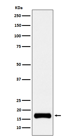
Anti-ISG15 Rabbit Monoclonal Antibody
- SPECIFICATION
- CITATIONS
- PROTOCOLS
- BACKGROUND

Application
| WB, IF, ICC |
|---|---|
| Primary Accession | P05161 |
| Host | Rabbit |
| Isotype | IgG |
| Reactivity | Human |
| Clonality | Monoclonal |
| Format | Liquid |
| Description | Anti-ISG15 Rabbit Monoclonal Antibody . Tested in WB, ICC/IF applications. This antibody reacts with Human. |
| Gene ID | 9636 |
|---|---|
| Other Names | Ubiquitin-like protein ISG15, Interferon-induced 15 kDa protein, Interferon-induced 17 kDa protein, IP17, Ubiquitin cross-reactive protein, hUCRP, ISG15 (HGNC:4053), G1P2, UCRP |
| Calculated MW | 17 kDa |
| Application Details | WB 1:500-1:2000 ICC/IF 1:50-1:200 |
| Contents | Rabbit IgG in phosphate buffered saline, pH 7.4, 150mM NaCl, 0.02% sodium azide and 50% glycerol, 0.4-0.5mg/ml BSA. |
| Clone Names | Clone: 21I77 |
| Immunogen | A synthesized peptide derived from human ISG15 |
| Purification | Affinity-chromatography |
| Storage | Store at -20°C for one year. For short term storage and frequent use, store at 4°C for up to one month. Avoid repeated freeze-thaw cycles. |
| Name | ISG15 (HGNC:4053) |
|---|---|
| Synonyms | G1P2, UCRP |
| Function | Ubiquitin-like protein which plays a key role in the innate immune response to viral infection either via its conjugation to a target protein (ISGylation) or via its action as a free or unconjugated protein (PubMed:27564865, PubMed:39465252). ISGylation involves a cascade of enzymatic reactions involving E1, E2, and E3 enzymes which catalyze the conjugation of ISG15 to a lysine residue in the target protein (PubMed:33727702). Its target proteins include IFIT1, MX1/MxA, PPM1B, UBE2L6, UBA7, CHMP5, CHMP2A, CHMP4B and CHMP6. Isgylation of the viral sensor IFIH1/MDA5 promotes IFIH1/MDA5 oligomerization and triggers activation of innate immunity against a range of viruses, including coronaviruses, flaviviruses and picornaviruses (PubMed:33727702). Can also isgylate: EIF2AK2/PKR which results in its activation, RIGI which inhibits its function in antiviral signaling response, EIF4E2 which enhances its cap structure-binding activity and translation-inhibition activity, UBE2N and UBE2E1 which negatively regulates their activity, IRF3 which inhibits its ubiquitination and degradation and FLNB which prevents its ability to interact with the upstream activators of the JNK cascade thereby inhibiting IFNA-induced JNK signaling. Exhibits antiviral activity towards both DNA and RNA viruses, including influenza A, HIV-1 and Ebola virus. Restricts HIV-1 and ebola virus via disruption of viral budding. Inhibits the ubiquitination of HIV-1 Gag and host TSG101 and disrupts their interaction, thereby preventing assembly and release of virions from infected cells. Inhibits Ebola virus budding mediated by the VP40 protein by disrupting ubiquitin ligase activity of NEDD4 and its ability to ubiquitinate VP40. ISGylates influenza A virus NS1 protein which causes a loss of function of the protein and the inhibition of virus replication. The secreted form of ISG15 can: induce natural killer cell proliferation, act as a chemotactic factor for neutrophils and act as a IFN-gamma-inducing cytokine playing an essential role in antimycobacterial immunity. The secreted form acts through the integrin ITGAL/ITGB2 receptor to initiate activation of SRC family tyrosine kinases including LYN, HCK and FGR which leads to secretion of IFNG and IL10; the interaction is mediated by ITGAL (PubMed:29100055). |
| Cellular Location | Cytoplasm. Secreted Note=Exists in three distinct states: free within the cell, released into the extracellular space, or conjugated to target proteins |
| Tissue Location | Detected in lymphoid cells, striated and smooth muscle, several epithelia and neurons. Expressed in neutrophils, monocytes and lymphocytes. Enhanced expression seen in pancreatic adenocarcinoma, endometrial cancer, and bladder cancer, as compared to non-cancerous tissue. In bladder cancer, the increase in expression exhibits a striking positive correlation with more advanced stages of the disease. |

Thousands of laboratories across the world have published research that depended on the performance of antibodies from Abcepta to advance their research. Check out links to articles that cite our products in major peer-reviewed journals, organized by research category.
info@abcepta.com, and receive a free "I Love Antibodies" mug.
Provided below are standard protocols that you may find useful for product applications.
If you have used an Abcepta product and would like to share how it has performed, please click on the "Submit Review" button and provide the requested information. Our staff will examine and post your review and contact you if needed.
If you have any additional inquiries please email technical services at tech@abcepta.com.


 Foundational characteristics of cancer include proliferation, angiogenesis, migration, evasion of apoptosis, and cellular immortality. Find key markers for these cellular processes and antibodies to detect them.
 The SUMOplot™ Analysis Program predicts and scores sumoylation sites in your protein. SUMOylation is a post-translational modification involved in various cellular processes, such as nuclear-cytosolic transport, transcriptional regulation, apoptosis, protein stability, response to stress, and progression through the cell cycle.
 The Autophagy Receptor Motif Plotter predicts and scores autophagy receptor binding sites in your protein. Identifying proteins connected to this pathway is critical to understanding the role of autophagy in physiological as well as pathological processes such as development, differentiation, neurodegenerative diseases, stress, infection, and cancer.


