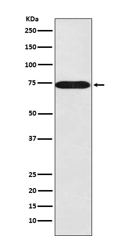
Anti-SNX1 Rabbit Monoclonal Antibody
- SPECIFICATION
- CITATIONS
- PROTOCOLS
- BACKGROUND

Application
| WB, IHC, IF, ICC, FC |
|---|---|
| Primary Accession | Q13596 |
| Host | Rabbit |
| Isotype | IgG |
| Reactivity | Rat, Human, Mouse |
| Clonality | Monoclonal |
| Format | Liquid |
| Description | Anti-SNX1 Rabbit Monoclonal Antibody . Tested in WB, IHC, ICC/IF, Flow Cytometry applications. This antibody reacts with Human, Mouse, Rat. |
| Gene ID | 6642 |
|---|---|
| Other Names | Sorting nexin-1, SNX1 |
| Calculated MW | 74 kDa |
| Application Details | WB 1:1000-1:5000 IHC 1:50-1:200 ICC/IF 1:50-1:200 FC 1:50 |
| Contents | Rabbit IgG in phosphate buffered saline, pH 7.4, 150mM NaCl, 0.02% sodium azide and 50% glycerol, 0.4-0.5mg/ml BSA. |
| Clone Names | Clone: 20S26 |
| Immunogen | A synthesized peptide derived from human SNX1 |
| Purification | Affinity-chromatography |
| Storage | Store at -20°C for one year. For short term storage and frequent use, store at 4°C for up to one month. Avoid repeated freeze-thaw cycles. |
| Name | SNX1 |
|---|---|
| Function | Involved in several stages of intracellular trafficking. Interacts with membranes containing phosphatidylinositol 3-phosphate (PtdIns(3P)) or phosphatidylinositol 3,5-bisphosphate (PtdIns(3,5)P2) (PubMed:12198132). Acts in part as component of the retromer membrane- deforming SNX-BAR subcomplex. The SNX-BAR retromer mediates retrograde transport of cargo proteins from endosomes to the trans-Golgi network (TGN) and is involved in endosome-to-plasma membrane transport for cargo protein recycling. The SNX-BAR subcomplex functions to deform the donor membrane into a tubular profile called endosome-to-TGN transport carrier (ETC) (Probable). Can sense membrane curvature and has in vitro vesicle-to-membrane remodeling activity (PubMed:19816406, PubMed:23085988). Involved in retrograde endosome-to-TGN transport of lysosomal enzyme receptors (IGF2R, M6PR and SORT1) and Shiginella dysenteria toxin stxB. Plays a role in targeting ligand-activated EGFR to the lysosomes for degradation after endocytosis from the cell surface and release from the Golgi (PubMed:12198132, PubMed:15498486, PubMed:17101778, PubMed:17550970, PubMed:18088323, PubMed:21040701). Involvement in retromer-independent endocytic trafficking of P2RY1 and lysosomal degradation of protease-activated receptor-1/F2R (PubMed:16407403, PubMed:20070609). Promotes KALRN- and RHOG-dependent but retromer-independent membrane remodeling such as lamellipodium formation; the function is dependent on GEF activity of KALRN (PubMed:20604901). Required for endocytosis of DRD5 upon agonist stimulation but not for basal receptor trafficking (PubMed:23152498). |
| Cellular Location | Endosome membrane; Peripheral membrane protein; Cytoplasmic side. Golgi apparatus, trans-Golgi network membrane; Peripheral membrane protein; Cytoplasmic side. Early endosome membrane; Peripheral membrane protein; Cytoplasmic side. Cell projection, lamellipodium. Note=Enriched on tubular elements of the early endosome membrane. Binds preferentially to highly curved membranes enriched in phosphatidylinositol 3-phosphate (PtdIns(3P)) or phosphatidylinositol 3,5-bisphosphate (PtdIns(3,5)P2) (PubMed:15498486). Colocalized with SORT1 to tubular endosomal membrane structures called endosome-to-TGN transport carriers (ETCs) which are budding from early endosome vacuoles just before maturing into late endosome vacuoles (PubMed:18088323). Colocalizes with DNAJC13 and Shiginella dysenteria toxin stxB on early endosomes (PubMed:19874558) Colocalized with F-actin at the leading edge of lamellipodia in a KALRN-dependent manner (PubMed:20604901). |

Thousands of laboratories across the world have published research that depended on the performance of antibodies from Abcepta to advance their research. Check out links to articles that cite our products in major peer-reviewed journals, organized by research category.
info@abcepta.com, and receive a free "I Love Antibodies" mug.
Provided below are standard protocols that you may find useful for product applications.
If you have used an Abcepta product and would like to share how it has performed, please click on the "Submit Review" button and provide the requested information. Our staff will examine and post your review and contact you if needed.
If you have any additional inquiries please email technical services at tech@abcepta.com.


 Foundational characteristics of cancer include proliferation, angiogenesis, migration, evasion of apoptosis, and cellular immortality. Find key markers for these cellular processes and antibodies to detect them.
 The SUMOplot™ Analysis Program predicts and scores sumoylation sites in your protein. SUMOylation is a post-translational modification involved in various cellular processes, such as nuclear-cytosolic transport, transcriptional regulation, apoptosis, protein stability, response to stress, and progression through the cell cycle.
 The Autophagy Receptor Motif Plotter predicts and scores autophagy receptor binding sites in your protein. Identifying proteins connected to this pathway is critical to understanding the role of autophagy in physiological as well as pathological processes such as development, differentiation, neurodegenerative diseases, stress, infection, and cancer.


