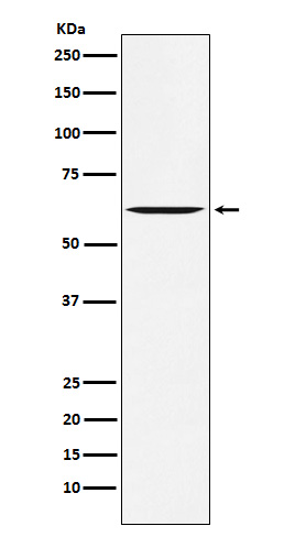
Anti-Yes1 Rabbit Monoclonal Antibody
- SPECIFICATION
- CITATIONS
- PROTOCOLS
- BACKGROUND

Application
| WB, IHC, IF, ICC, IP |
|---|---|
| Primary Accession | P07947 |
| Host | Rabbit |
| Isotype | IgG |
| Reactivity | Rat, Human, Mouse |
| Clonality | Monoclonal |
| Format | Liquid |
| Description | Anti-Yes1 Rabbit Monoclonal Antibody . Tested in WB, IHC, ICC/IF, IP applications. This antibody reacts with Human, Mouse, Rat. |
| Gene ID | 7525 |
|---|---|
| Other Names | Tyrosine-protein kinase Yes, 2.7.10.2, Proto-oncogene c-Yes, p61-Yes, YES1, YES |
| Calculated MW | 60801 Da |
| Application Details | WB 1:500-1:2000 IHC 1:50-1:200 ICC/IF 1:50-1:200 IP 1:50 |
| Contents | Rabbit IgG in phosphate buffered saline, pH 7.4, 150mM NaCl, 0.02% sodium azide and 50% glycerol, 0.4-0.5mg/ml BSA. |
| Clone Names | Clone: 19Y44 |
| Immunogen | A synthesized peptide derived from human Yes1 |
| Purification | Affinity-chromatography |
| Storage | Store at -20°C for one year. For short term storage and frequent use, store at 4°C for up to one month. Avoid repeated freeze-thaw cycles. |
| Name | YES1 |
|---|---|
| Synonyms | YES |
| Function | Non-receptor protein tyrosine kinase that is involved in the regulation of cell growth and survival, apoptosis, cell-cell adhesion, cytoskeleton remodeling, and differentiation. Stimulation by receptor tyrosine kinases (RTKs) including EGFR, PDGFR, CSF1R and FGFR leads to recruitment of YES1 to the phosphorylated receptor, and activation and phosphorylation of downstream substrates. Upon EGFR activation, promotes the phosphorylation of PARD3 to favor epithelial tight junction assembly. Participates in the phosphorylation of specific junctional components such as CTNND1 by stimulating the FYN and FER tyrosine kinases at cell-cell contacts. Upon T-cell stimulation by CXCL12, phosphorylates collapsin response mediator protein 2/DPYSL2 and induces T-cell migration. Participates in CD95L/FASLG signaling pathway and mediates AKT-mediated cell migration. Plays a role in cell cycle progression by phosphorylating the cyclin-dependent kinase 4/CDK4 thus regulating the G1 phase. Also involved in G2/M progression and cytokinesis. Catalyzes phosphorylation of organic cation transporter OCT2 which induces its transport activity (PubMed:26979622). |
| Cellular Location | Cell membrane. Cytoplasm, cytoskeleton, microtubule organizing center, centrosome. Cytoplasm, cytosol. Cell junction {ECO:0000250|UniProtKB:Q28923}. Note=Newly synthesized protein initially accumulates in the Golgi region and traffics to the plasma membrane through the exocytic pathway. Localized to small puncta throughout the cytoplasm and cell membrane when in the presence of SNAIL1 (By similarity). {ECO:0000250|UniProtKB:Q28923} |
| Tissue Location | Expressed in the epithelial cells of renal proximal tubules and stomach as well as hematopoietic cells in the bone marrow and spleen in the fetal tissues. In adult, expressed in epithelial cells of the renal proximal tubules and present in keratinocytes in the basal epidermal layer of epidermis. |

Thousands of laboratories across the world have published research that depended on the performance of antibodies from Abcepta to advance their research. Check out links to articles that cite our products in major peer-reviewed journals, organized by research category.
info@abcepta.com, and receive a free "I Love Antibodies" mug.
Provided below are standard protocols that you may find useful for product applications.
If you have used an Abcepta product and would like to share how it has performed, please click on the "Submit Review" button and provide the requested information. Our staff will examine and post your review and contact you if needed.
If you have any additional inquiries please email technical services at tech@abcepta.com.


 Foundational characteristics of cancer include proliferation, angiogenesis, migration, evasion of apoptosis, and cellular immortality. Find key markers for these cellular processes and antibodies to detect them.
 The SUMOplot™ Analysis Program predicts and scores sumoylation sites in your protein. SUMOylation is a post-translational modification involved in various cellular processes, such as nuclear-cytosolic transport, transcriptional regulation, apoptosis, protein stability, response to stress, and progression through the cell cycle.
 The Autophagy Receptor Motif Plotter predicts and scores autophagy receptor binding sites in your protein. Identifying proteins connected to this pathway is critical to understanding the role of autophagy in physiological as well as pathological processes such as development, differentiation, neurodegenerative diseases, stress, infection, and cancer.


