


 Foundational characteristics of cancer include proliferation, angiogenesis, migration, evasion of apoptosis, and cellular immortality. Find key markers for these cellular processes and antibodies to detect them.
 The SUMOplot™ Analysis Program predicts and scores sumoylation sites in your protein. SUMOylation is a post-translational modification involved in various cellular processes, such as nuclear-cytosolic transport, transcriptional regulation, apoptosis, protein stability, response to stress, and progression through the cell cycle.
 The Autophagy Receptor Motif Plotter predicts and scores autophagy receptor binding sites in your protein. Identifying proteins connected to this pathway is critical to understanding the role of autophagy in physiological as well as pathological processes such as development, differentiation, neurodegenerative diseases, stress, infection, and cancer.
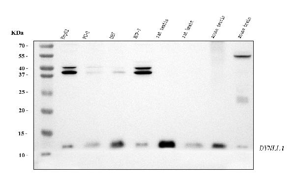
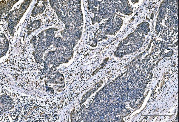
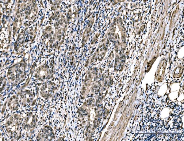
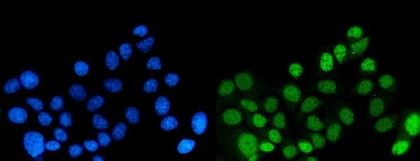
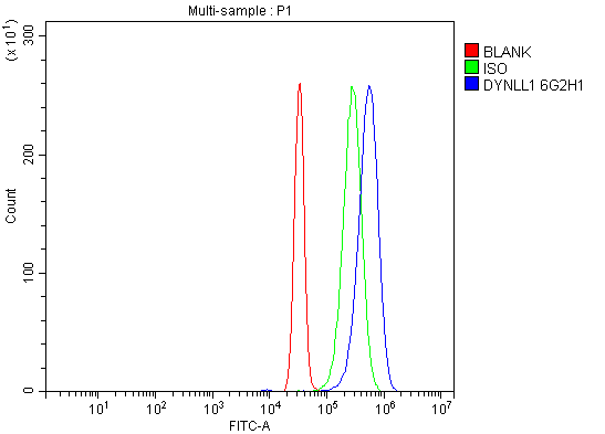
Anti-DYNLL1/PIN Antibody Picoband™ (monoclonal, 6G2H1)
- SPECIFICATION
- CITATIONS
- PROTOCOLS
- BACKGROUND

Application
| WB, IHC, IF, ICC, FC |
|---|---|
| Primary Accession | P63167 |
| Host | Mouse |
| Isotype | Mouse IgG2b |
| Reactivity | Rat, Human, Mouse |
| Clonality | Monoclonal |
| Format | Lyophilized |
| Description | Anti-DYNLL1/PIN Antibody Picoband™ (monoclonal, 6G2H1) . Tested in Flow Cytometry, IF, IHC, ICC, WB applications. This antibody reacts with Human, Mouse, Rat. |
| Reconstitution | Adding 0.2 ml of distilled water will yield a concentration of 500 µg/ml. |
| Gene ID | 8655 |
|---|---|
| Other Names | Dynein light chain 1, cytoplasmic, 8 kDa dynein light chain, DLC8, Dynein light chain LC8-type 1, Protein inhibitor of neuronal nitric oxide synthase, PIN, DYNLL1 {ECO:0000303|Ref.9, ECO:0000312|HGNC:HGNC:15476} |
| Calculated MW | 12 kDa |
| Application Details | Western blot, 0.25-0.5 µg/ml, Human, Mouse, Rat Immunohistochemistry(Paraffin-embedded Section), 2-5 µg/ml, Human Immunocytochemistry/Immunofluorescence, 5 µg/ml, Human Flow Cytometry, 1-3 µg/1x10^6 cells, Human |
| Contents | Each vial contains 4 mg Trehalose, 0.9 mg NaCl and 0.2 mg Na2HPO4. |
| Clone Names | Clone: 6G2H1 |
| Immunogen | E.coli-derived human DYNLL1/PIN recombinant protein (Position: M1-G89). |
| Purification | Immunogen affinity purified. |
| Storage | At -20°C for one year from date of receipt. After reconstitution, at 4°C for one month. It can also be aliquotted and stored frozen at -20°C for six months. Avoid repeated freezing and thawing. |
| Name | DYNLL1 {ECO:0000303|Ref.9, ECO:0000312|HGNC:HGNC:15476} |
|---|---|
| Function | Acts as one of several non-catalytic accessory components of the cytoplasmic dynein 1 complex that are thought to be involved in linking dynein to cargos and to adapter proteins that regulate dynein function (By similarity). Cytoplasmic dynein 1 acts as a motor for the intracellular retrograde motility of vesicles and organelles along microtubules (By similarity). May play a role in changing or maintaining the spatial distribution of cytoskeletal structures (By similarity). In addition to its role in cytoskeleton and transport, acts as a protein-protein adapter, which inhibits and/or sequesters target proteins (PubMed:10198631, PubMed:15193260, PubMed:15891768, PubMed:16684779, PubMed:30464262, PubMed:37696958). Involved in the response to DNA damage by acting as a key regulator of DNA end resection: when phosphorylated at Ser-88, recruited to DNA double- strand breaks (DSBs) by TP53BP1 and acts by disrupting MRE11 dimerization, thereby inhibiting DNA end resection (PubMed:30464262, PubMed:37696958). In a subset of DSBs, DYNLL1 remains unphosphorylated and promotes the recruitment of the Shieldin complex (PubMed:37696958). Binds and inhibits the catalytic activity of neuronal nitric oxide synthase/NOS1 (By similarity). Promotes transactivation functions of ESR1 and plays a role in the nuclear localization of ESR1 (PubMed:15891768, PubMed:16684779). Regulates apoptotic activities of BCL2L11 by sequestering it to microtubules (PubMed:10198631, PubMed:15193260). Upon apoptotic stimuli the BCL2L11-DYNLL1 complex dissociates from cytoplasmic dynein and translocates to mitochondria and sequesters BCL2 thus neutralizing its antiapoptotic activity (PubMed:10198631, PubMed:15193260). |
| Cellular Location | Cytoplasm, cytoskeleton, microtubule organizing center, centrosome. Chromosome. Cytoplasm, cytoskeleton. Nucleus Mitochondrion. Note=Upon induction of apoptosis translocates together with BCL2L11 to mitochondria (PubMed:18084006). Recruited to DNA double-strand breaks (DSBs) by TP53BP1 when phosphorylated at Ser-88 (PubMed:37696958) |
| Tissue Location | Ubiquitous (PubMed:8628263). Expressed in testis (PubMed:22965910). |

Thousands of laboratories across the world have published research that depended on the performance of antibodies from Abcepta to advance their research. Check out links to articles that cite our products in major peer-reviewed journals, organized by research category.
info@abcepta.com, and receive a free "I Love Antibodies" mug.

Provided below are standard protocols that you may find useful for product applications.
Background
Dynein light chain 1, cytoplasmic is a protein that in humans is encoded by the DYNLL1 gene. Cytoplasmic dyneins are large enzyme complexes with a molecular mass of about 1,200 kD. They contain two force-producing heads formed primarily from dynein heavy chains, and stalks linking the heads to a basal domain, which contains a varying number of accessory intermediate chains. The complex is involved in intracellular transport and motility. The protein described in this record is a light chain and exists as part of this complex but also physically interacts with and inhibits the activity of neuronal nitric oxide synthase. Binding of this protein destabilizes the neuronal nitric oxide synthase dimer, a conformation necessary for activity, and it may regulate numerous biologic processes through its effects on nitric oxide synthase activity. Alternate transcriptional splice variants have been characterized.
If you have used an Abcepta product and would like to share how it has performed, please click on the "Submit Review" button and provide the requested information. Our staff will examine and post your review and contact you if needed.
If you have any additional inquiries please email technical services at tech@abcepta.com.
Ordering Information
Other Products
Shipping Information
