


 Foundational characteristics of cancer include proliferation, angiogenesis, migration, evasion of apoptosis, and cellular immortality. Find key markers for these cellular processes and antibodies to detect them.
 The SUMOplot™ Analysis Program predicts and scores sumoylation sites in your protein. SUMOylation is a post-translational modification involved in various cellular processes, such as nuclear-cytosolic transport, transcriptional regulation, apoptosis, protein stability, response to stress, and progression through the cell cycle.
 The Autophagy Receptor Motif Plotter predicts and scores autophagy receptor binding sites in your protein. Identifying proteins connected to this pathway is critical to understanding the role of autophagy in physiological as well as pathological processes such as development, differentiation, neurodegenerative diseases, stress, infection, and cancer.
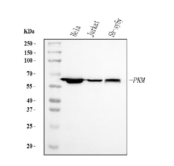
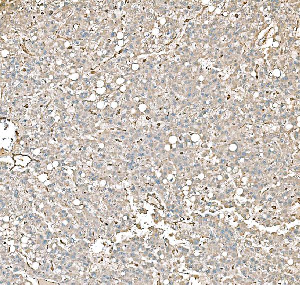
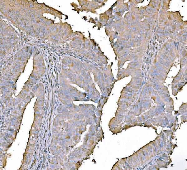
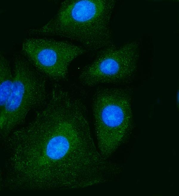
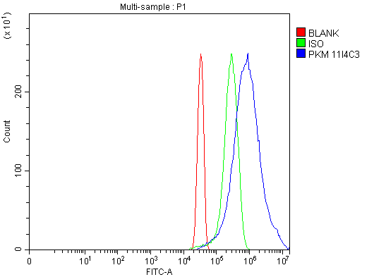
Anti-PKM2 Antibody Picoband™ (monoclonal, 11I4C3)
- SPECIFICATION
- CITATIONS
- PROTOCOLS
- BACKGROUND

Application
| WB, IHC, IF, ICC, FC |
|---|---|
| Primary Accession | P14618 |
| Host | Mouse |
| Isotype | Mouse IgG2b |
| Reactivity | Human |
| Clonality | Monoclonal |
| Format | Lyophilized |
| Description | Anti-PKM2 Antibody Picoband™ (monoclonal, 11I4C3) . Tested in Flow Cytometry, IF, IHC, ICC, WB applications. This antibody reacts with Human. |
| Reconstitution | Adding 0.2 ml of distilled water will yield a concentration of 500 µg/ml. |
| Gene ID | 5315 |
|---|---|
| Other Names | Pyruvate kinase PKM, 2.7.1.40, Cytosolic thyroid hormone-binding protein, CTHBP, Opa-interacting protein 3, OIP-3, Pyruvate kinase 2/3, Pyruvate kinase muscle isozyme, Threonine-protein kinase PKM2, 2.7.11.1, Thyroid hormone-binding protein 1, THBP1, Tumor M2-PK, Tyrosine-protein kinase PKM2, 2.7.10.2, p58, PKM, OIP3 {ECO:0000303|PubMed:9466265}, PK2, PK3, PKM2 |
| Calculated MW | 60 kDa |
| Application Details | Western blot, 0.25-0.5 µg/ml, Human Immunohistochemistry(Paraffin-embedded Section), 2-5 µg/ml, Human Immunocytochemistry/Immunofluorescence, 5 µg/ml, Human Flow Cytometry, 1-3 µg/1x10^6 cells, Human |
| Contents | Each vial contains 4 mg Trehalose, 0.9 mg NaCl and 0.2 mg Na2HPO4. |
| Clone Names | Clone: 11I4C3 |
| Immunogen | A synthetic peptide corresponding to a sequence at the N-terminus of human PKM2, different from the related mouse sequence by five amino acids, and from the related rat sequence by four amino acids. |
| Purification | Immunogen affinity purified. |
| Storage | At -20°C for one year from date of receipt. After reconstitution, at 4°C for one month. It can also be aliquotted and stored frozen at -20°C for six months. Avoid repeated freezing and thawing. |
| Name | PKM |
|---|---|
| Synonyms | OIP3 {ECO:0000303|PubMed:9466265}, PK2, |
| Function | Catalyzes the final rate-limiting step of glycolysis by mediating the transfer of a phosphoryl group from phosphoenolpyruvate (PEP) to ADP, generating ATP (PubMed:15996096, PubMed:1854723, PubMed:20847263). The ratio between the highly active tetrameric form and nearly inactive dimeric form determines whether glucose carbons are channeled to biosynthetic processes or used for glycolytic ATP production (PubMed:15996096, PubMed:1854723, PubMed:20847263). The transition between the 2 forms contributes to the control of glycolysis and is important for tumor cell proliferation and survival (PubMed:15996096, PubMed:1854723, PubMed:20847263). |
| Cellular Location | [Isoform M2]: Cytoplasm. Nucleus Note=Translocates to the nucleus in response to various signals, such as EGF receptor activation or apoptotic stimuli (PubMed:17308100, PubMed:22056988, PubMed:24120661). Nuclear translocation is promoted by acetylation by EP300 (PubMed:24120661). Deacetylation by SIRT6 promotes its nuclear export in a process dependent of XPO4, thereby suppressing its ability to activate transcription and promote tumorigenesis (PubMed:26787900). |
| Tissue Location | [Isoform M2]: Specifically expressed in proliferating cells, such as embryonic stem cells, embryonic carcinoma cells, as well as cancer cells. |

Thousands of laboratories across the world have published research that depended on the performance of antibodies from Abcepta to advance their research. Check out links to articles that cite our products in major peer-reviewed journals, organized by research category.
info@abcepta.com, and receive a free "I Love Antibodies" mug.

Provided below are standard protocols that you may find useful for product applications.
Background
PKM (Pyruvate Kinase, Muscle), also known as PK3 or PKM2, is an enzyme that in humans is encoded by the PKM gene. The activity of pyruvate kinase subtype M2 is increased by fructose 1, 6-bisphosphate (Fru-1, 6-P2). By in situ hybridization, Popescu and Cheng (1990) mapped the THBP1 gene to 15q24-q25. Ashizawa et al. (1991) manipulated the intracellular Fru-1, 6-P2 concentration in several mammalian cell lines, including human, by varying the glucose concentration in the media. Using a novel proteomic screen for phosphotyrosine-binding proteins, Christofk et al. (2008) observed that PKM2 binds directly and selectively to tyrosine-phosphorylated peptides.
If you have used an Abcepta product and would like to share how it has performed, please click on the "Submit Review" button and provide the requested information. Our staff will examine and post your review and contact you if needed.
If you have any additional inquiries please email technical services at tech@abcepta.com.
Ordering Information
Other Products
Shipping Information
